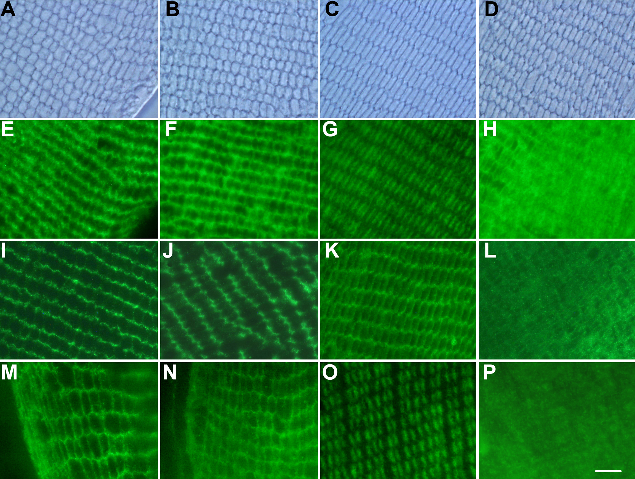
Figure 5. Localization of filensin and
phakinin in the 10-week-old rat lens. The localization of filensin in
10-week-old rat lenses was examined by phase-contrast microscopy (A-D)
and fluorescence immunochemistry using antibodies directed against the
filensin rod domain (E-H), filensin outer tail domain (I-L),
and phakinin (M-P). (A, E, I, and M)
shallow cortex of the Wistar lens; (B, F, J, and
N) shallow cortex of the pre-cataract SCR lens; (C, G,
K, and O) deep cortex of the Wistar lens; (D, H,
L and P) deep cortex of the SCR lens. The anti-filensin
rod domain antibody localized to the membrane lining regions in the
shallow cortices of Wistar and pre-cataract SCR lenses (E and F)
and to the central region of the cytoplasm in the deep cortex of the
Wistar lens (G). The anti-filensin rod domain antibody exhibited
a diffuse staining pattern in the deep cortex of the cataract SCR lens (H).
The anti-filensin outer tail domain antibody localized to the membrane
lining region of the shallow cortex of the Wistar (I) and
pre-cataract SCR lens (J) as well as the deep cortex of the
Wistar lens (K). This antibody exhibited a diffuse staining
pattern in the deep cortex of the pre-cataract SCR lens. The
localization of phakinin (M-P) was similar to that of the
filensin rod domain (E-H). Scale bar, 10 µm.