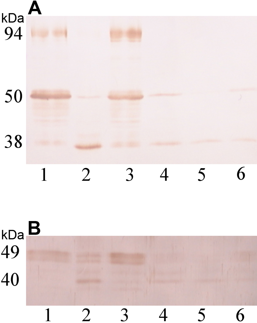
Figure 2. Western blot analysis of
filensin and phakinin in the 10-week-old rat lens. SDS–PAGE and western
blot analysis of the cortical and nuclear regions of lenses from
10-week-old Wistar rats, pre-cataract SCRs, and normal SCRs using an
anti-filensin antibody (A) and an anti-phakinin antibody (B)
are shown. Ten micrograms of protein were loaded in each lane (Lane 1,
cortex of Wistar lens; lane 2, cortex of pre-cataract SCR lens; lane 3,
cortex of normal SCR lens; lane 4, nucleus of Wistar lens; lane 5,
nucleus of pre-cataract SCR lens; lane 6, nucleus of normal SCR lens).
The levels of filensin and phakinin in the nuclear regions were lower
in 10-week-old lenses than in 6-week-old lenses. The 94 kDa form of
filensin was absent from the cortex of the pre-cataract SCR lens. The
50 kDa form of filensin was decreased and the 38 kDa form was increased
in the cortices of pre-cataract lenses as compared to Wistar lenses.