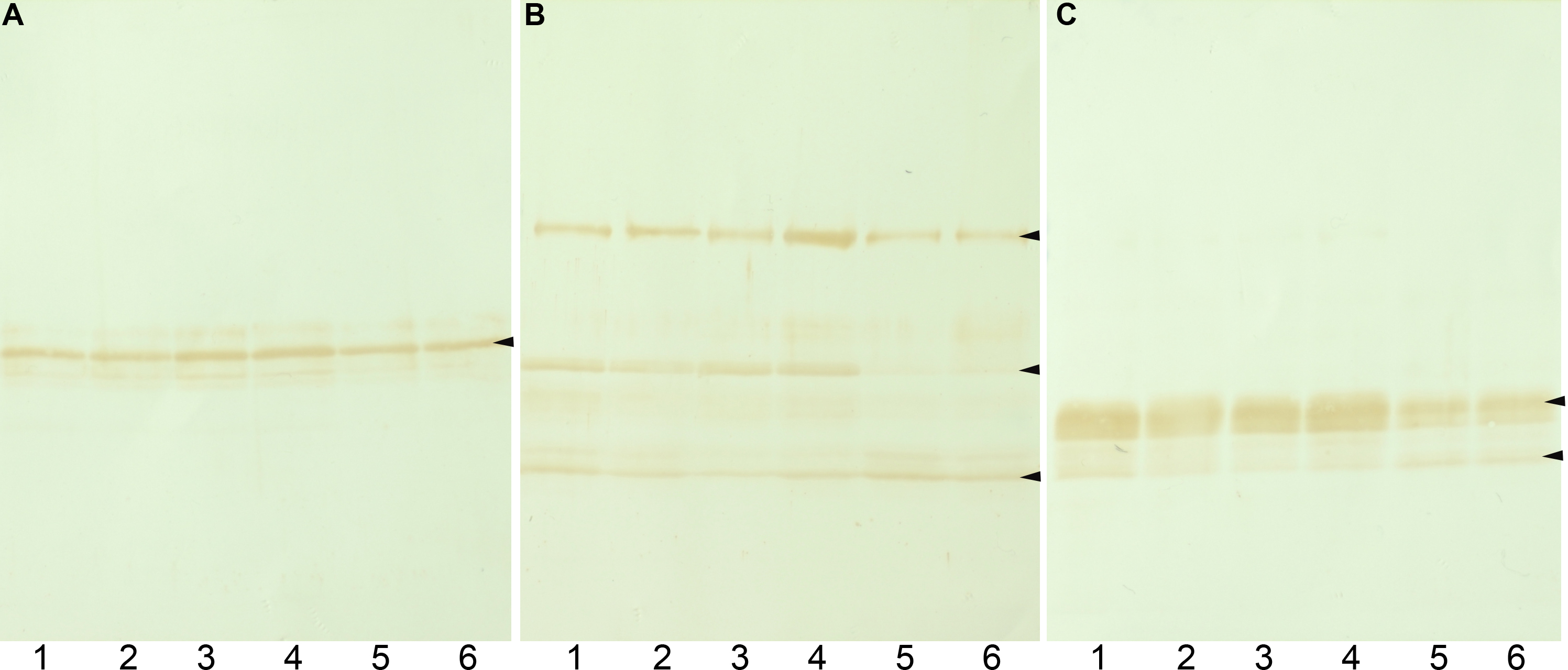
Figure 1. Western blot analysis of
intermediate filament proteins in the lens. Western blot analysis of
vimentin (A), filensin (B), and phakinin (C) in
the lenses of 10-week-old Wistar rats (lanes 1 and 2), normal SCRs
(lanes 3 and 4), and pre-cataract SCRs (lanes 5 and 6) is shown. The
levels of the 50 kDa form filensin and phakinin in pre-cataract lenses
were lower than in control lenses.