Figure 7. Persistent remodeling of the choroid-retinal pigment epithelium (RPE) interface over a dorsoventral gradient. Visualization
Top: Quantitative gray-scale images of glutamine signals displayed as density. Visualization Bottom: Serial section stained
with toluidine blue. Up arrows, RPE; o, disrupted rod outer segments in the corner of the eye-shaped survivor zone photoreceptor
outer segment layer; H, Haller’s layer of the choroid. Scale: The top panel is over 5 mm long and the bottom image is 0.410
mm wide. The enlarged view shows the complete loss of the choriocapillaris and RPE in the LD zone and complete retention (arrows)
in the survivor zone. The large vessel Haller’s layer of the choroid directly abuts the remnant retina in the (LD zone dotted
line). ONH, optic nerve head. Sample metadata: SD Rat, age at LX 60 d, animal #VO 01–72 SDDRLD3–1AM-2WK-3L, left eye, 3 h
LX starting at 1AM, harvested at 14 days pLX, bloc code 6563, slide code 3322.
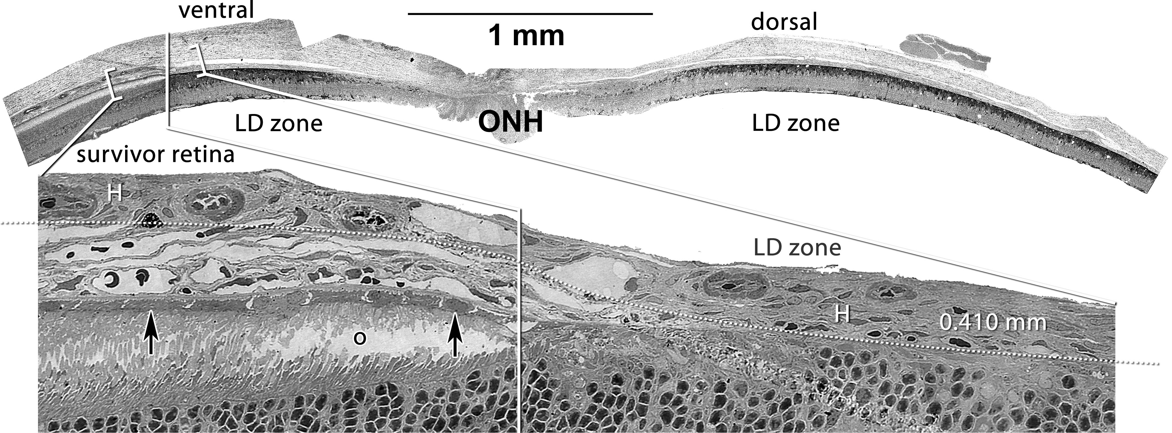
 Figure 7 of
Marc, Mol Vis 2008; 14:782-805.
Figure 7 of
Marc, Mol Vis 2008; 14:782-805.