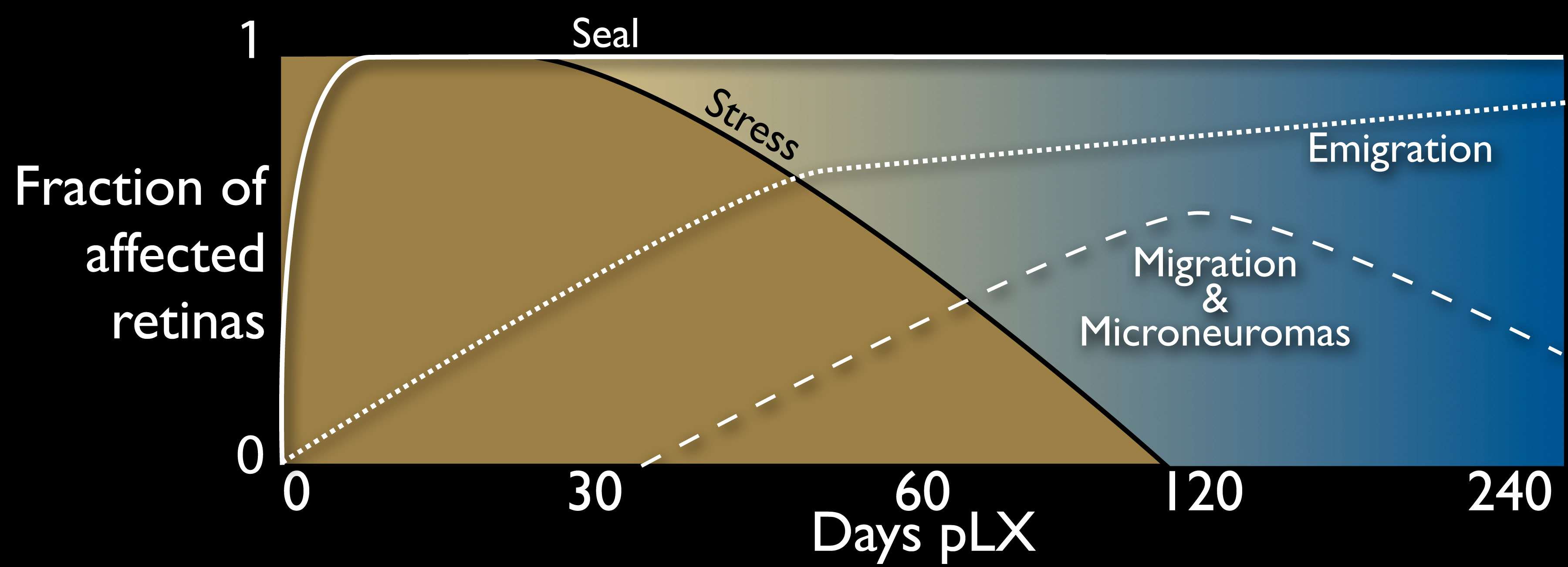
Figure 19. Temporal profile of remodeling
in light induced retinal damage. A schematic of the fraction of retinas
displaying remodeling attributes (see
Table 3) displayed on an
exponential time scale. Immediately after light exposure, all retinas
show massive stress signals, but by pLX 120 these are no longer
evident. Müller cell seals form rapidly after light exposure and
persist. Breakdown of the seal can be detected by pLX 14 and increases
with time. Classic remodeling phenomena such as migration and
microneuromas are evident by pLX60 but decline after emigration and
retinal decimation become dominant.
 Figure 19 of Marc, Mol Vis 2008; 14:782-805.