Figure 1. A summary of the key attributes
of light-induced retinal damage across the albino rat retina. A:
The dorsoventral gradient of alterations manifested over time. At late
stages, normal retina is restricted to ventral retina near the ora
terminalis. The bulk of intact retina is (B) survivor retina
with shortened photoreceptors. At early stages, zones of (C)
stressed retina with stressed photoreceptors are large but then
contract to a thin rim near the light damage (LD) margin. Also at early
stages, transient buckling (D) appears at the junction of intact
retina and the LD margin (E) as photoreceptors continue to
produce outer segment material, but retinal pigmented epithelium (RPE)
cells are compromised. LD retina forms (F) remodeling zones with
internal revisions such as neuronal migration, microneuromas and fluid
channel formation; (G) emigration sites with aggressive
migration of retina cells into the remnant choroid, leaving behind
decimated zones with severely depleted retinas. Abbreviations: BCs
represents bipolar cells, ACs represents amacrine cells, GCs represents
ganglion cells, CC represents choriocapillaris, MCs represents Müller
cells.
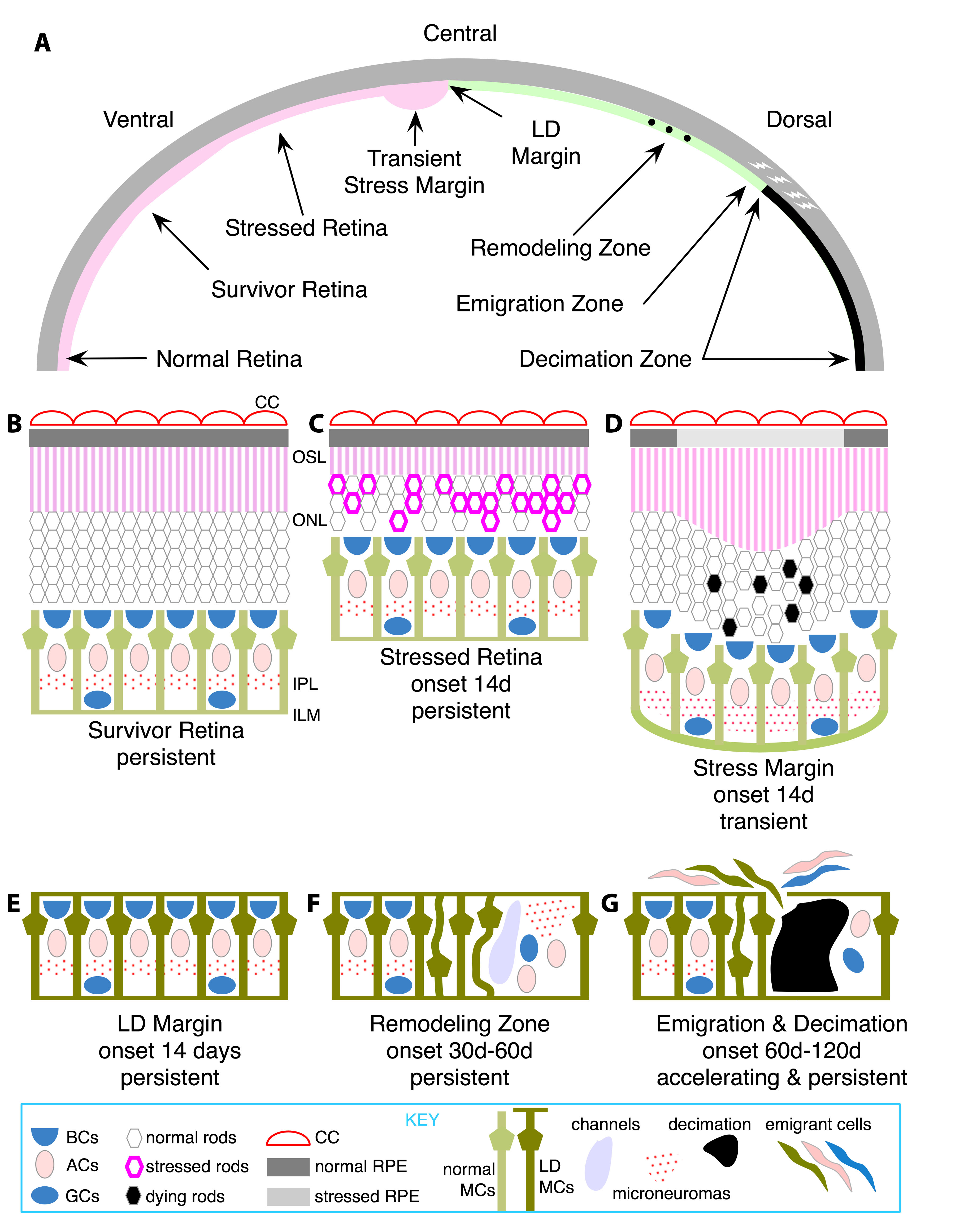
 Figure 1 of Marc, Mol Vis 2008; 14:782-805.