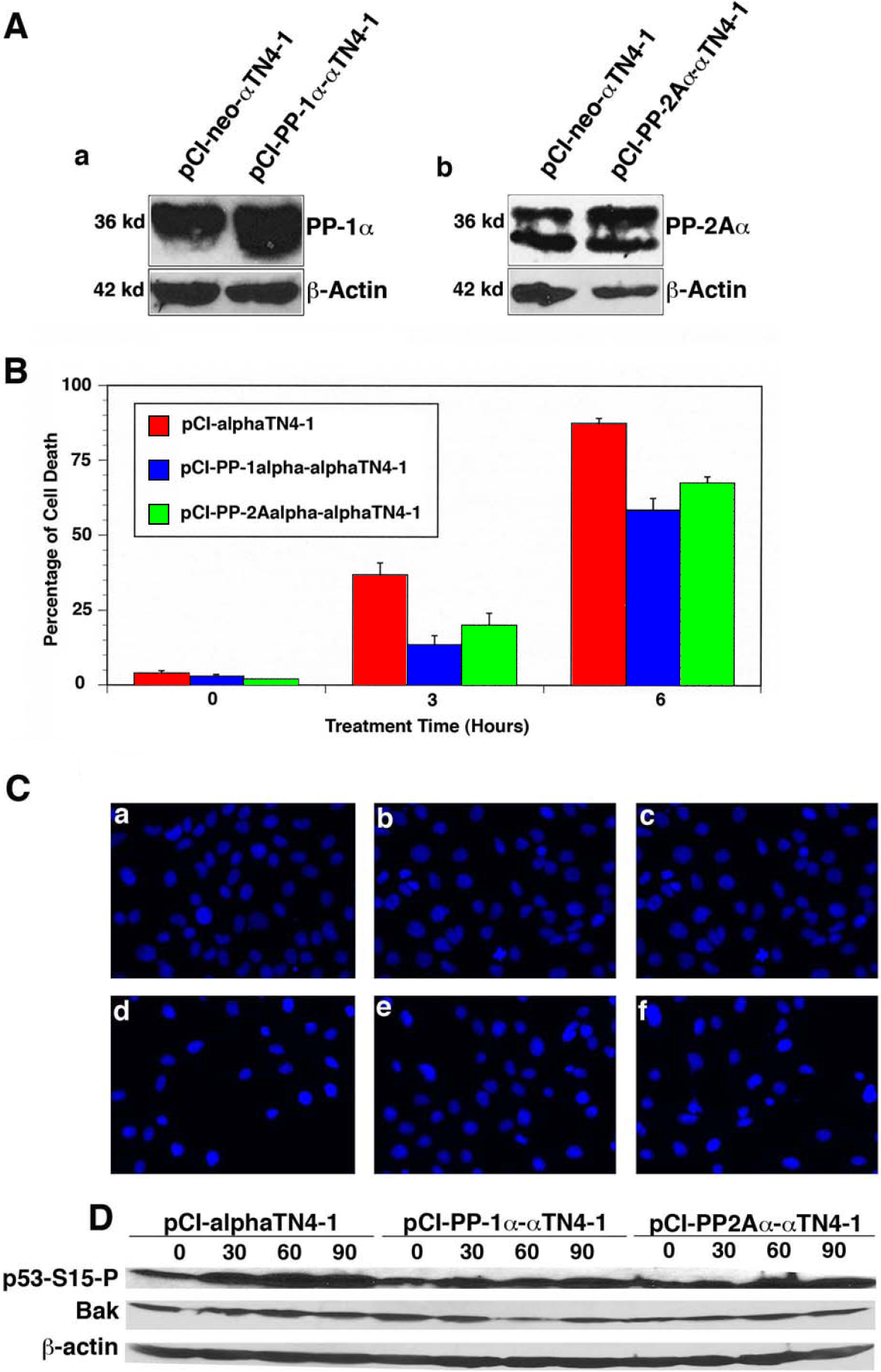
Figure 8. PP-1 and PP-2A protect lens
epithelial cells from oxidative stress-induced apoptosis.
A:
western blot analysis confirms the overexpression of PP-1α (a) and
PP2Aα (b) in αTN4–1 cells. The stable clones expressing the pCI-neo
vector, pCI-PP-1α, or pCI-PP-2Aα were obtained through G418 screen (400
µg/ml). The expression levels of PP-1α and PP-2Aα were determined by
western blot analysis using 100 µg of total protein extracted from
pCI-neo or PP-1α-tranfected αTN4–1 cells (a) and from pCI-neo or
PP-2Aα-transfected αTN4–1 cells (b).
B: Results of the MTT
assay is shown in the chart. The MTT assay is described in the Methods
section. Note that PP-1α displayed a stronger ability against oxidative
stress-induced cell death than PP-2Aα did.
C: Hoechst staining
analysis of the pCI-neotransfected cells (a, d), PP-1α-tranfected
αTN4–1 cells (b, e), or PP-2Aα-transfected αTN4–1 cells (c, f) without
treatment (a, b, c) or treated by 85–95 µM H2O2 (d, e, f) for 3.5 h is
pictured. Hoechst staining was conducted as previously described [
14,
17]. Note that after
treatment, the apoptotic cells became either dissociated from the
culture plate (thus leaving empty space in the culture dish) or
condensed.
D: Western blot analysis of the p53
hyperphosphorylation at Ser-15 and Bak expression. The western blot
analysis was conducted as described before [
23]. Note that in both PP-1α and
PP-2Aα-transfected cells, hydrogen peroxide-induced
hyperphosphorylation of p53 at Ser-53 and Bak upregulation were
obviously attenuated.
 Figure 8 of Liu, Mol Vis 2008; 14:762-773.