Figure 5. Western blot for AQP5 in
keratoconus and non-keratoconus epithelial cells. The photograph of a
western blot identifies two bands of AQP5. Twenty-seven and 31 kDa
corresponds to the non-glycosylated protein and glycosylated form of
the AQP5 protein, respectively. Lane A corresponds to a non-KC sample;
lane B corresponds to a KC sample. GAPDH protein is shown below as an
internal control.
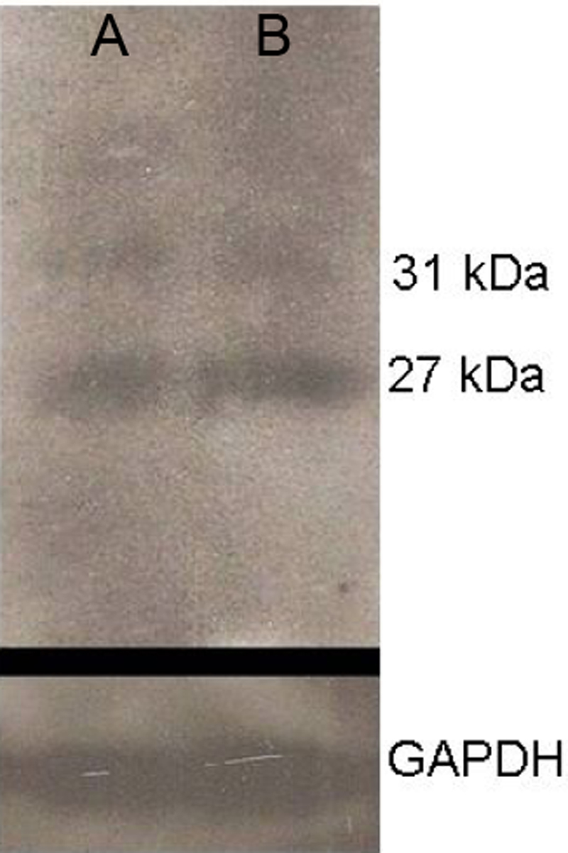
 Figure 5 of Garfias, Mol Vis 2008; 14:756-761.