Figure 3. AQP5 immunohistochemistry from keratoconus and non-keratoconus corneal tissue. A: The immunopositive reaction to AQP5 from a KC sample is shown as a brown signal in the epithelial cells. B: Non-KC tissue demonstrates a similar immunopositive AQP5 reaction. Note disruptions in Bowman’s layer in A. b, Bowman’s layer; e, corneal epithelium; s, stroma (Representative assay of 29 KC and 14 non-KC samples). Bar=100 µm.
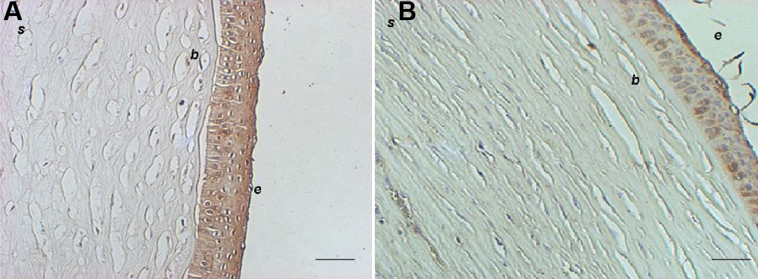
 Figure 3 of
Garfias, Mol Vis 2008; 14:756-761.
Figure 3 of
Garfias, Mol Vis 2008; 14:756-761.