Figure 2. Fundus pictures of affected patients showed that the cup/disc ratios of the affected patients were very high. A: The picture displays the left optic nerve head of subject IV:1 in Family A. B and C: The last two pictures presents the left (B) and right (C) optic nerve head of subject V:14 of Family B. Note nerve fiber layer defect, notching, and excavation in all three fundus
pictures. The disc asymmetry was shown between the two eyes of V:14 in Family B (B, C).
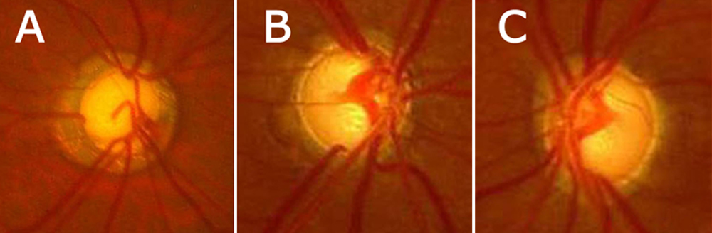
 Figure 2 of
Lin, Mol Vis 2008; 14:739-744.
Figure 2 of
Lin, Mol Vis 2008; 14:739-744.