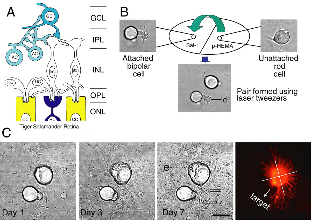
Figure 1. Movement and culture of cells grouped as pairs with optical tweezers.
A: Schematic organization of the salamander retina based on Lasansky [
9,
10] and Wong-Riley [
11]. Rod (RC) and cone cells (CC) connect with second-order neurons, horizontal (HC) and bipolar cells (BC), which in turn connect
with third-order neurons, amacrine (AC) and ganglion cells (GC). Normal interactions for photoreceptors, therefore, are exclusively
with second-order neurons.
B: Creating cell pairs with laser tweezers. A rod cell on the poly-HEMA side of the culture dish (upper right, see Methods for
more details) is selected, trapped in the laser beam and transported to the Sal-1 side where it is placed less than 10 µm
away from a bipolar cell (upper left) to form a pair (lower middle). The bipolar cell was identified by the presence of a
thick Landolt club (lc). The photoreceptor was identified by the presence of an ellipsoid best seen in part
C (e).
C: Analysis of growth. Growth was followed for one week in culture. Photoreceptor identification, initially made by the presence
of an ellipsoid (e) and cell shape, was confirmed by immunopositive staining for rod opsin (far right). This rod photoreceptor
is attracted to the bipolar cell. At day 1, a thick lamellar process forms from the photoreceptor cell, growing toward neurites
emanating from the bipolar Landolt club. By day 7, long neuritic processes with varicosities (arrows) were present; some processes
were contacting the dendrites of the target cell. Quantitative analysis of growth in the quadrants toward and away from the
target (far right) verified that, in addition to multiple contacts, there were more varicosities toward the target. Scale
bar equals 20 µm.
 Figure 1 of
Clarke, Mol Vis 2008; 14:706-720.
Figure 1 of
Clarke, Mol Vis 2008; 14:706-720.