Figure 1. Splicing assays of wild-type and mutant PRPF31 in transfected HEK 293T cells. A: Assays using RHO intron 3 minigene splicing template (RHO-MG). As a positive control, cells were transfected with the splicing template only (RHO-MG only). Cells transfected with WT PRPF31 only gave no products (negative control). Marker for the unspliced product was generated by amplification directly from the
plasmid construct using the same primers. B: Bar graphs show the splicing efficiencies, derived from the relative band strengths
for cells expressing untagged and His-tagged PRPF31. Error bars indicate the standard error of means derived from four separate
determinations. Double asterisks (**) indicate that the reduced splicing efficiencies of mutant splicing factor compared to
the positive and negative controls are statistically significant (p<0.01). C: Assays using full length RHO (FL-RHO) splicing template. Analysis of all four introns indicates 100% splicing efficiency. No unspliced transcript was detected
in cDNA from human retina. D: Assays using GNAT1 template.
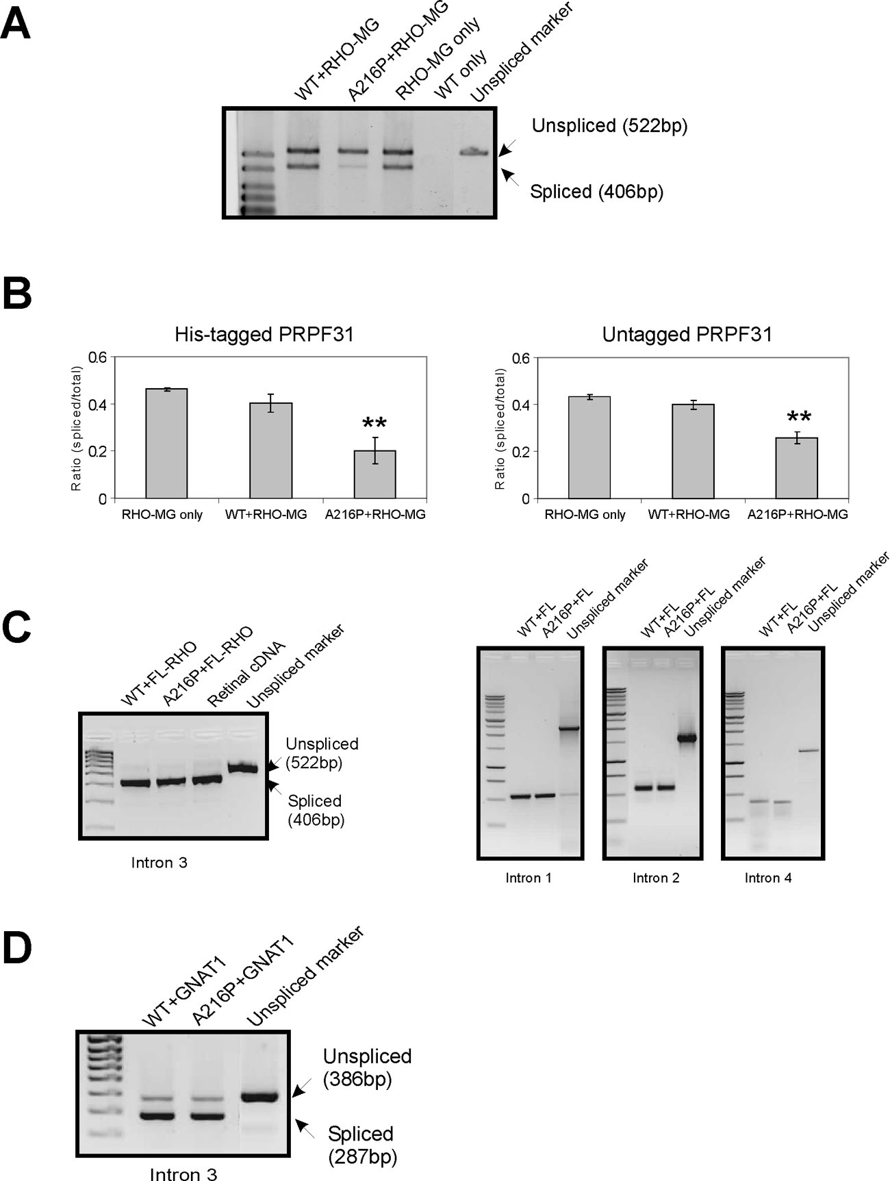
 Figure 1 of
Wilkie, Mol Vis 2008; 14:683-690.
Figure 1 of
Wilkie, Mol Vis 2008; 14:683-690.