Figure 3. The c.1495+2_1495+3insT mutation. A: Shown are nucleotide sequence traces of the boundary between TULP1 exon and intron14 in a non-carrier individual (wt), an individual heterozygote for the c.1495+2_1495+3insT mutation (het),
and an affected individual homozygote for the mutant allele (mut). The exon-intron boundary is marked. B: A mismatch-primer restriction–based assay used to screen control DNA samples for the c.1495+2_1495+3insT mutation. The reverse
PCR primer included one mismatched base, which, in combination with the mutant allele, but not with the WT allele, created
a DrdI restriction site. A 286 bp PCR product derived from the WT allele was not digested by DrdI, while in the presence of
the MUT two restriction fragments of 251 and 35 bp were obtained. Digestion of a PCR product derived from a heterozygote for
the mutation (HET) yielded three fragments (286, 251, and 35 bp). The 35 bp fragment is not visible.
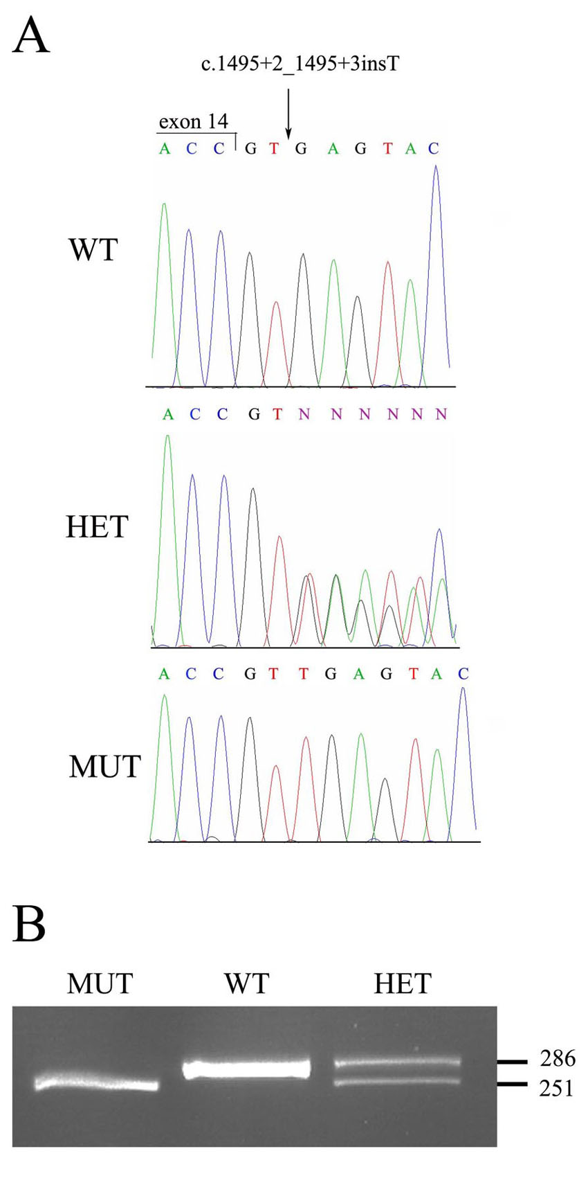
 Figure 3 of
Abbasi, Mol Vis 2008; 14:675-681.
Figure 3 of
Abbasi, Mol Vis 2008; 14:675-681.