Figure 2. Fundus photographs of individual IV-18 (right eye). The photographs show vascular attenuation, peripheral bone spicule pigmentation,
accompanied by salt and pepper-like changes, and signs of maculopathy, including edema and a yellow perifoveal ring.
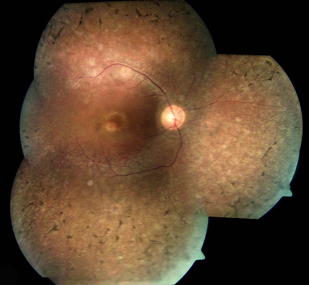
 Figure 2 of
Abbasi, Mol Vis 2008; 14:675-681.
Figure 2 of
Abbasi, Mol Vis 2008; 14:675-681.