![]() Figure 4 of
Ishino, Mol Vis 2008;
14:61-70.
Figure 4 of
Ishino, Mol Vis 2008;
14:61-70.
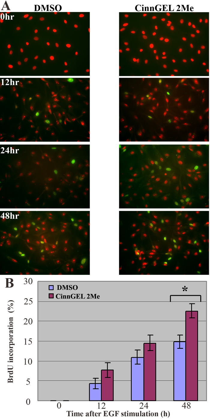
Figure 4. Effect of PTP1B inhibitor on cell cycle entry in response to EGF stimulation
Subconfluent HCEC were pre-incubated for 1 h in medium containing either DMSO alone or 25 μM of the PTP1B inhibitor, CinnGEL 2Me, diluted in DMSO. Following this pre-incubation step, 25 ng/ml EGF was added to all cultures. Samples were taken for BrdU staining at 0, 12, 24, or 48 h after EGF addition. A: Representative images showing BrdU staining (green) in subconfluent HCEC from a 72 year-old donor incubated in either DMSO alone or in CinnGEL 2Me PTP1B inhibitor. Cultures were counterstained with propidium iodide (PI) to reveal all nuclei (red). Final magnification: 400X. B: Bar graph shows the average percent of BrdU-positive HCEC at each time point. Bars represent SEM. The asterisk indicates statistical significance at p=0.019.