![]() Figure 2 of
Ishino, Mol Vis 2008;
14:61-70.
Figure 2 of
Ishino, Mol Vis 2008;
14:61-70.
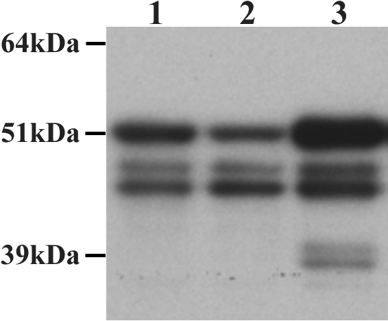
Figure 2. Western blot detection of PTP1B protein bands in HCEC
Confluent Passage-2 HCEC were cultured from a newborn (lane 1) and a 74-year-old donor (lane 2) and processed for western blot detection of PTP1B. A commercially prepared whole cell lysate of SW480 cells was used as a positive control (lane 3). Blots showed that the two HCEC samples yielded a band at approximately 50 kDa, indicating the presence of full-length PTP1B, as well as two additional bands of approximately 48 kDa and 46 kDa, possibly representing truncated forms. The same three bands were observed in the SW480 positive control sample, as well as lower molecular weight bands around 39-37 kDa that may result from non-specific proteolytic cleavage. Note the variability in density of the three PTP1B-positive bands within the two HCEC samples.