![]() Figure 1 of
Ishino, Mol Vis 2008;
14:61-70.
Figure 1 of
Ishino, Mol Vis 2008;
14:61-70.
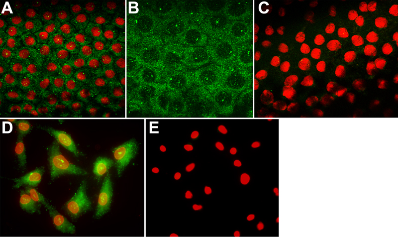
Figure 1. Representative images of PTP1B localization in ex vivo human corneal endothelium and in cultured HCEC
A: Representative image of PTP1B (green) staining in the endothelium of a cornea obtained from a 59-year-old donor. PTP1B is localized in a punctate pattern mainly within the cytoplasm. Some punctate staining is also visible in nuclei (red). Note that larger dots of intense stain can be observed scattered within the tissue or on individual cells. This appears to be due to non-specific antibody deposition, although all antibodies were centrifuged at high-speed before use. B: Higher magnification image of HCEC in ex vivo corneal tissue showing more detail of the PTP1B localization pattern. C: Negative control of ex vivo tissue in which corneas were incubated with secondary antibody alone. Image is an overlay from both the FITC and rhodamine channels. D: PTP1B staining in subconfluent HCEC cultured from a 55-year-old donor results in a similar punctate pattern in the cytoplasm and nucleus. E: Negative control in which cells were incubated with secondary antibody only. Image is an overlay from both the FITC and rhodamine channels. green: PTP1B. red: Propidium iodide. Original magnification for A, C, D, and E: 40X. Original magnification for B: 100X.