Figure 5. Protein levels of DNA repair
enzymes are decreased in aged retinal pigment epithelium and choroid. A:
Immunoblots of the DNA repair enzymes, OGG1 and MYH, are shown. Actin
was used as internal reference. Each group of three bands represents
protein extracts from both retinal pigment epithelium (RPE) and choroid
of three different animals. B: The differences in expression
levels were determined by scanning gels and determining the integrated
areas of the bands using Image-J software. Data are expressed as
normalized ratios to actin. There were significant decreases in aged
RPE and choroid of OGG1 (p=0.02, n=3) and MYH (p=0.001, n=3), compared
to young RPE and choroid.
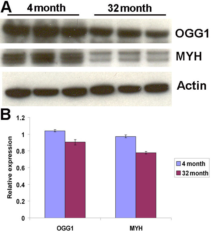
 Figure 5 of Wang, Mol Vis 2008; 14:644-651.