Figure 3. Increased levels of deleted
mitochondrial DNA in aged retinal pigment epithelium and choroid.
Measurements of levels of the PCR products of non-deleted (A,
not damaged) and deleted (B, damaged) mitochondrial DNA (mtDNA)
normalized by total mtDNA normalized by total mtDNA were done using the
PicoGreen reagent. Each PCR reaction started with 10 ng of genomic DNA
(nuclear and mitochondrial) from rat retinal pigment epithelium (RPE)
and choroid as a template. There was no difference in the non-deleted
mtDNA at 18 month (p=0.02, n=5), 24 month (p=0.06, n=5), and 32 month
(p=0.70, n=5), compared with the 4 month group. However, there was a
significant increase in deleted mtDNA at 18 month (p=0.003, n=5), 24
month (p<0.0001, n=5), and 32 month (p<0.0001, n=5), as compared
with the 4 month group. Values are the mean±SEM.
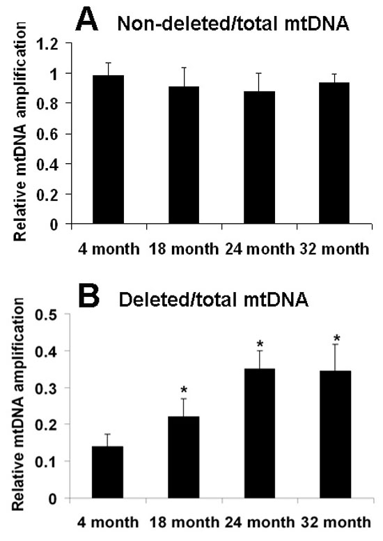
 Figure 3 of Wang, Mol Vis 2008; 14:644-651.