Figure 5. Immunodetection of α3, α5, αv, and β1 in materials obtained after precipitation of 92–1, Mel202, FM55P, and IGR-39 cell extracts with phaseolus vulgaris agglutinin
bound to agarose. One mg of the cell extracts were incubated overnight with phaseolus vulgaris agglutinin (PHA-L) immobilized
on cross-linked 4% beaded agarose. Glycoproteins were released from the complexes by boiling in electrophoresis sample buffer
before being subjected to 10% SDS–PAGE. Following separation, the proteins were blotted onto PVDF membrane. After being blocked
the blots were incubated with one of the following antibodies specific for different integrin subunits: α3, α5, αv, and β1. Next, the membranes were incubated with the secondary antibodies either alkaline phosphatase conjugated goat anti-rabbit
IgG (for α3, α5, αv, integrin subunits) or alkaline phosphatase coupled goat anti-mouse IgG (for β1 integrin subunit). Visualization of immunoreactive proteins was achieved with the use of 4-nitroblue-tetrazolium salt/5-bromo-4-chloro-3-indolylophosphate
solution. Lane S shows position of molecular weight markers.
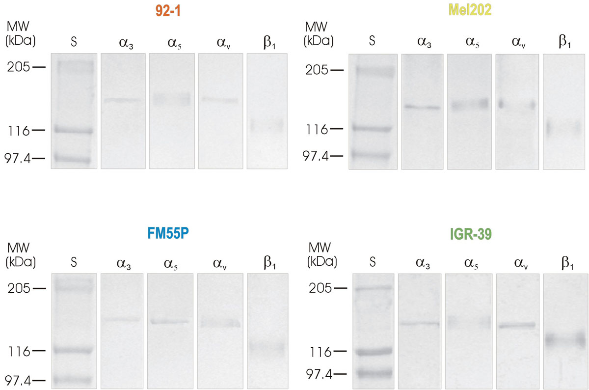
 Figure 5 of
Przybyło, Mol Vis 2008; 14:625-636.
Figure 5 of
Przybyło, Mol Vis 2008; 14:625-636.