Figure 5. Reactive gliosis is not detected following intracameral inoculation. Neuroretinal sonicate were collected out to day 24 and
assessed for glial fibrillary acidic protein (GFAP) expression via western blot analysis (n of 3 neuroretina time-point).
A representative western blot from day 8 demonstrates increased GFAP expression observed in positive control mice only. A: Averaged relative densities of respective GFAP bands (53 kDa) normalized to alpha-actin (42 kDa) loading control is summarized
in the bar graph. B: Levels of GFAP expression in normal eyes are not presented (0.22 – 0.54 relative expression units, n of 4). Asterisk (*)
indicates p value = 0.01, as calculated by ANOVA.
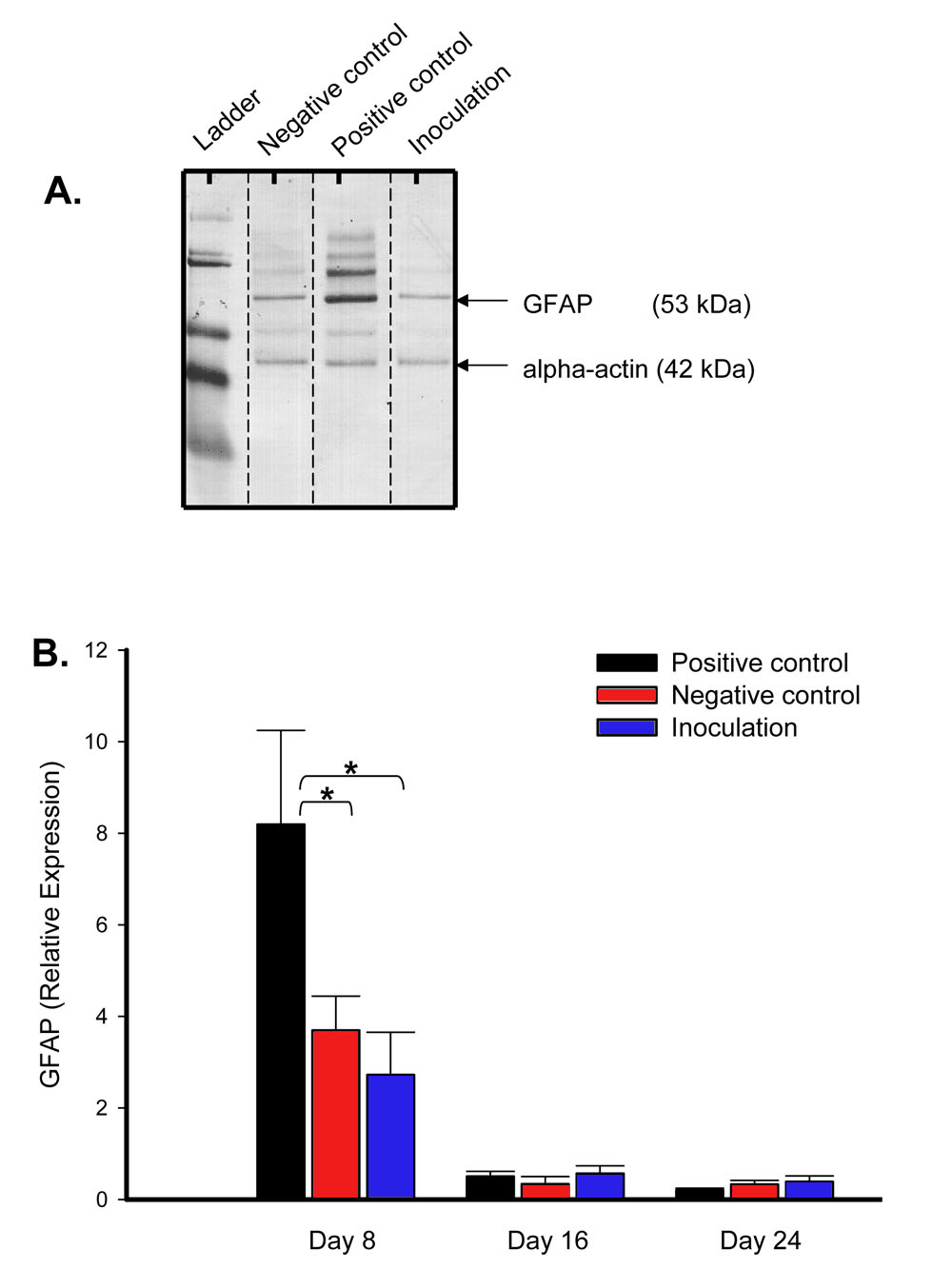
 Figure 5 of
Saban, Mol Vis 2008; 14:615-624.
Figure 5 of
Saban, Mol Vis 2008; 14:615-624.