Figure 3. Histopathology of anterior segment tissues following intracameral inoculation. Eyes were histologically examined at various
time-points out to 24 days following intracameral inoculation with allosplenocytes (n of 3 per micrograph). Micrographs (A,B) represent the histopathology observed in negative control mice eight and 16 days post sham operation. High-magnification
image demonstrates marginal scarring observed in these mice (A, inset). Micrographs (C,D) represent the histopathology observed in positive control mice eight and 16 days post anterior lens puncture. High-magnification
image (D) demonstrates inflammatory infiltrate in host anterior chamber (arrows). Micrographs (E,F) represent the histopathology observed eight and 16 days post intracameral inoculation with allosplenocytes. High magnification
image (E, inset) shows inflammatory infiltrate in host posterior cornea, which resolved by day 16 (F).
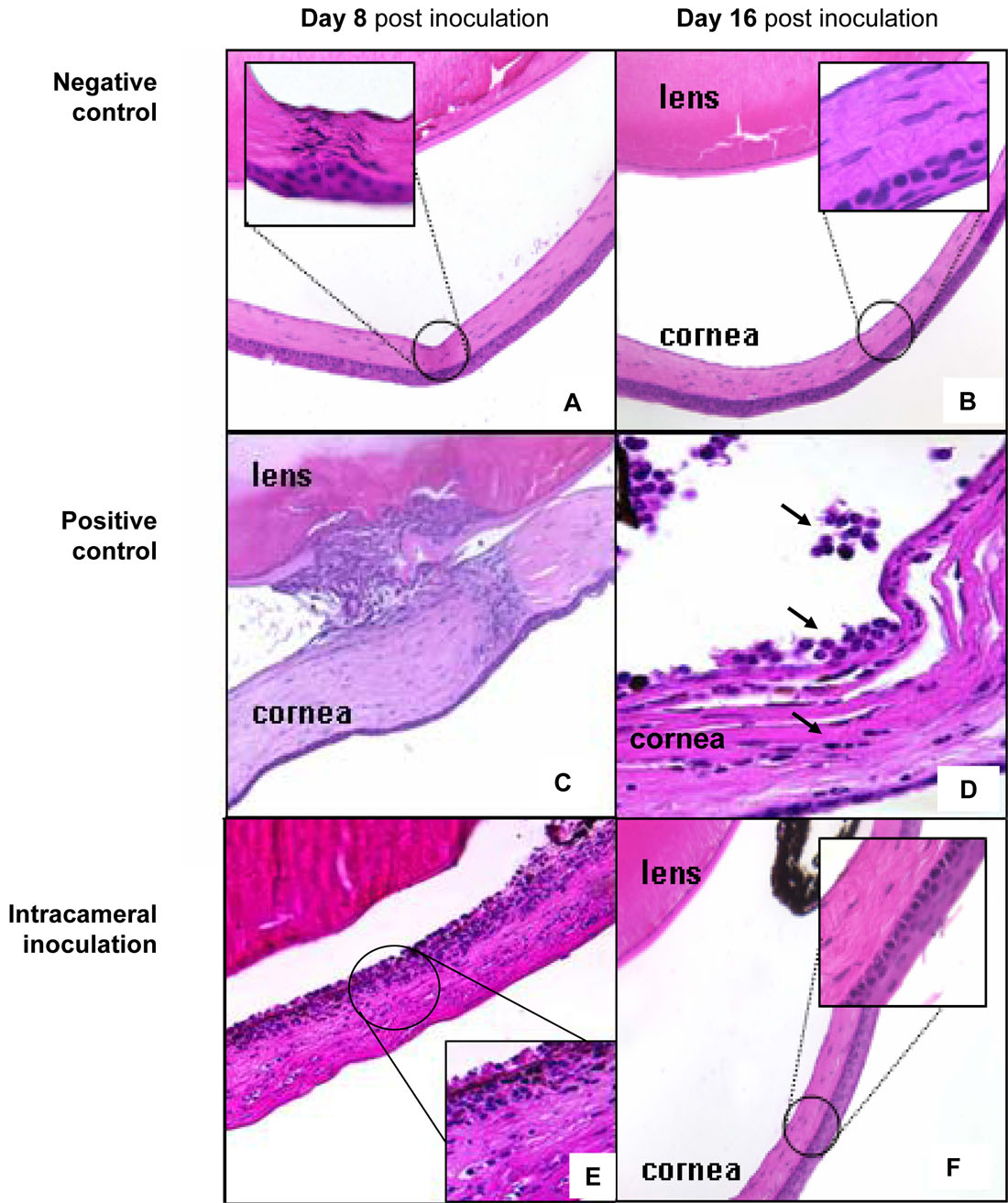
 Figure 3 of
Saban, Mol Vis 2008; 14:615-624.
Figure 3 of
Saban, Mol Vis 2008; 14:615-624.