Figure 2. Allosplenocytes are rapidly cleared from host anterior chamber following intracameral inoculation. BALB/c eyes were enucleated
at 3 to 24 h post intracameral inoculation (at least n=2 per micrograph) with enhanced green fluorescent protein (eGFP)+ allosplenocytes
(green). Some sections were counterstained with DAPI (blue) for epifluorescence microscopy. Representative high-magnification
images demonstrate a reduced presence of eGFP(+) observed in the central anterior chamber (AC) from 3 h (A) to 6 h (B) post inoculation. Representative low-magnification images demonstrate the few eGFP(+) cells observed in the central AC at
9 h (C), while eGFP(+) cells were abundant in the iridocorneal angles (D), and similarly observed at 12 h (E). Few eGFP(+) cells were also observed trafficking through the iris/ciliary body (D, inset). Host AC were absent of eGFP(+) cells by 24 h, with the exception of few cells and cell fragments remaining in some of the
inoculated mice (F).
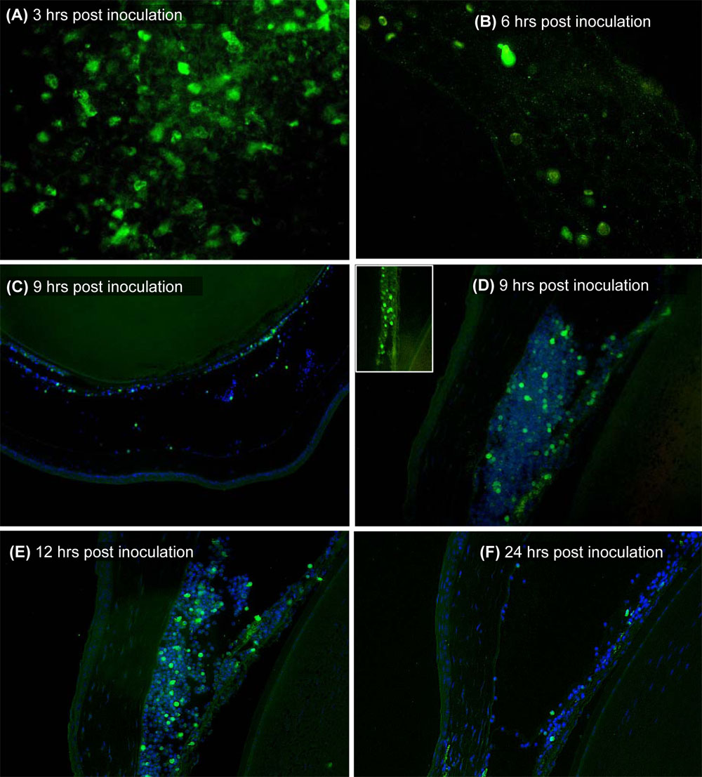
 Figure 2 of
Saban, Mol Vis 2008; 14:615-624.
Figure 2 of
Saban, Mol Vis 2008; 14:615-624.