Figure 5. Expression of Myc-E2F3a and E2F5 proteins. Immunohistochemistry was used to assay expression of the E2F3a and E2F5 transgenic
proteins in non-transgenic FVB (A, C) and the E2F3a/E2F5 double transgenic lenses (B, D). Using anti-Myc antibody, there is no green staining in FVB lens (A). By comparison, green nuclear staining is present in the transgenic lens fiber cells (B) indicative of Myc-tagged E2F3a expression. When antibody against E2F5 was used, endogenous E2F5 in FVB lens fiber cells
was detected as a weak green nuclear staining (C). A stronger signal was present in the double transgenic fiber cell nuclei (D). Scale bars=500 μm.
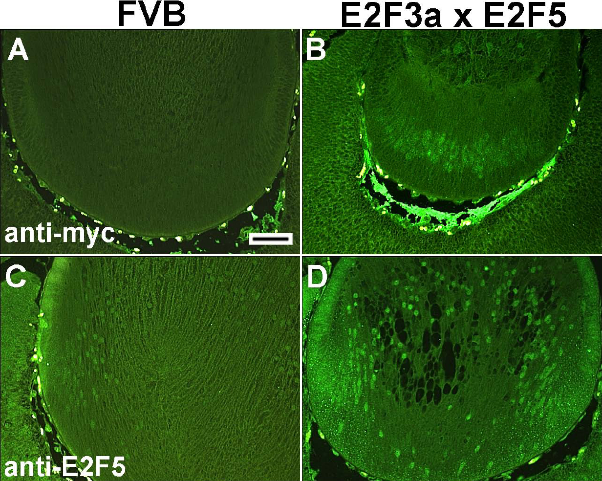
 Figure 5 of
Chen, Mol Vis 2008; 14:602-614.
Figure 5 of
Chen, Mol Vis 2008; 14:602-614.