Figure 3. Mutation in the SOX2 activation domain. Genomic DNA from affected patients, unaffected relatives, and healthy controls are amplified by PCR. PCR
products are separated by electrophoresis in an 8% polyacrylamide gel for detection of heteroduplexes and homoduplexes. Genomic
DNA samples are then sequenced for detection of sequence variants. A: Heteroduplexes detected by CSGE is shown. Lanes C1 and C2 represent the healthy controls; Lane 54A represents the proband;
Lane 54B represents the mother of the proband; Lane 54C represents the father of the proband; and Lane 54H represents the
affected sibling of the proband. The additional slower band in patient 54A may represent a conformational intermediate formed
during gel electrophoresis, resulting in a different mobility compared to homoduplexes and the triplet. B: Sequence analysis shows two alleles in the proband 54A, the affected sibling 54H, and the clinically normal mother 54B. One
allele is the wild type sequence and the other one is single nucleotide deletion at c.549delC. The father 54C is homozygous
for the wild type allele. C: The single nucleotide deletion at c.549delC results in a frame shift mutation and premature termination at 19 amino acids
downstream of the deletion site (p.Pro184ArgfsX19). The mutation is predicted to delete part of the C-terminal activation
domain and to produce a truncated peptide.
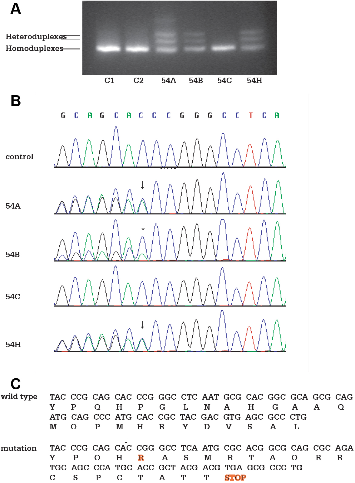
 Figure 3 of
Zhou, Mol Vis 2008; 14:583-592.
Figure 3 of
Zhou, Mol Vis 2008; 14:583-592.