Figure 2. Mutation in the SOX2 HMG domain. Genomic DNA from affected patients, unaffected relatives, and healthy controls are amplified by PCR. PCR products
are separated by electrophoresis in an 8% polyacrylamide gel for detection of heteroduplexes and homoduplexes. Genomic DNA
samples are then sequenced for detection of sequence variants. A: Heteroduplexes detected by CSGE is shown. Lanes C1 and C2 represent the healthy controls; Lane 40A represents the proband;
Lane 40B represents the mother of the proband; and Lane 40C is the father of the proband. Sample from patient 40A shows heteroduplex
comparing to homoduplex observed in samples from the parents (40B and 40C) and normal controls (C1 and C2). B: Single nucleotide substitution at c.310 G>T of the coding sequence is identified in patient 40A, but not in the parents (40B
and 40C) and normal controls (C1 and C2). C: Mutation at c.310 G>T results in a nonsense amino acid change at p. Glu104X as illustrated. HMG=high mobility group.
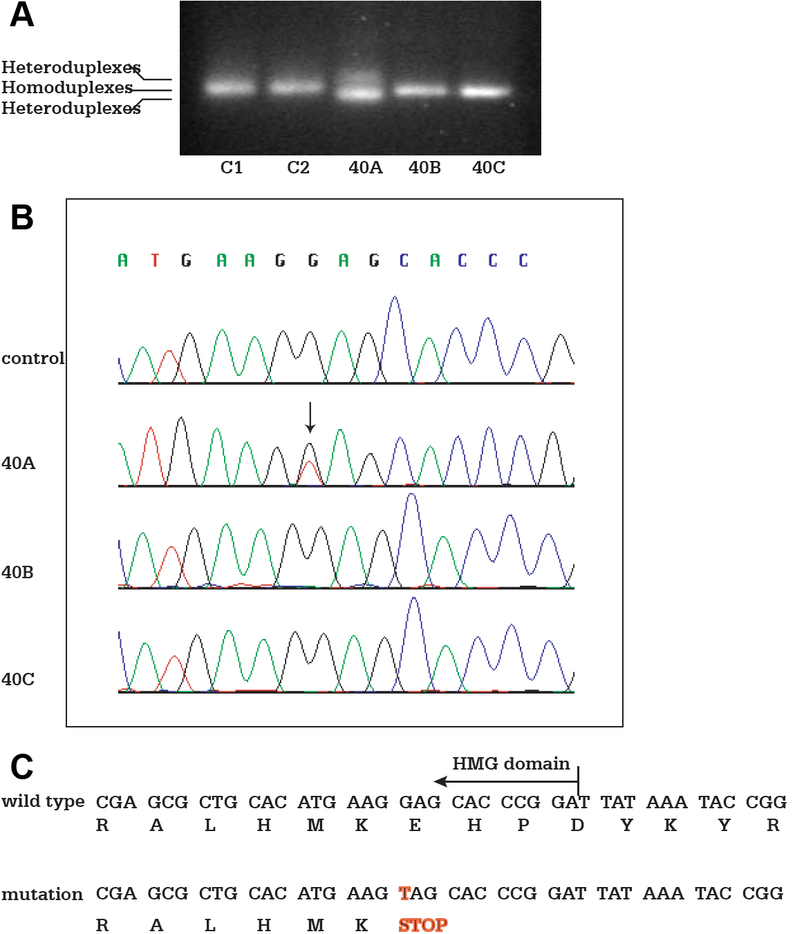
 Figure 2 of
Zhou, Mol Vis 2008; 14:583-592.
Figure 2 of
Zhou, Mol Vis 2008; 14:583-592.