Figure 4. Membranes of MLBs. As shown in the transmission electron micrographs, MLBs from age-related nuclear cataracts often appear
circular (B, C, and F) with an occasional doublet formation (E). MLBs are covered with multiple layers of lipid membranes, although these membranes can barely be visualized at low magnification
(A) in this donor lens. With proper tilt (25°-40°) and high magnification (D, from the inset box in A), the 10 membranes appear multilamellar with thin spacing (4.5 nm).
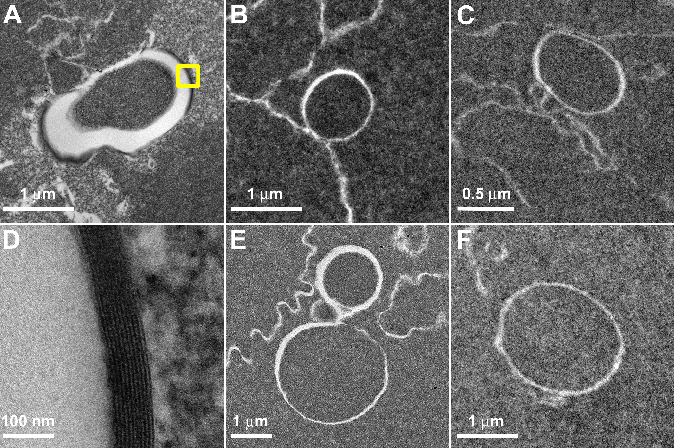
 Figure 4 of
Gilliland, Mol Vis 2008; 14:572-582.
Figure 4 of
Gilliland, Mol Vis 2008; 14:572-582.