Figure 1. Methods. Transparent lenses and age-related cataractous nuclei were immersion-fixed and sectioned with a Vibratome (A). The 160 µm thick sections were stained with the lipophilic dye, Fast DiI, washed with ethanol, and transferred to glass
slides with Permount and coverslips. For analysis, sections at or near the center of the lens were used so that all developmental
regions could be identified (C), including the embryonic nucleus (en), fetal nucleus (fn), juvenile nucleus (jn), adult nucleus (an), and cortex (c). Samples
were examined with laser scanning confocal microscopy (D). Individual volumes of tissue from each Vibratome section were visualized. The red line in (B) indicates an example of one focal plane that was used.
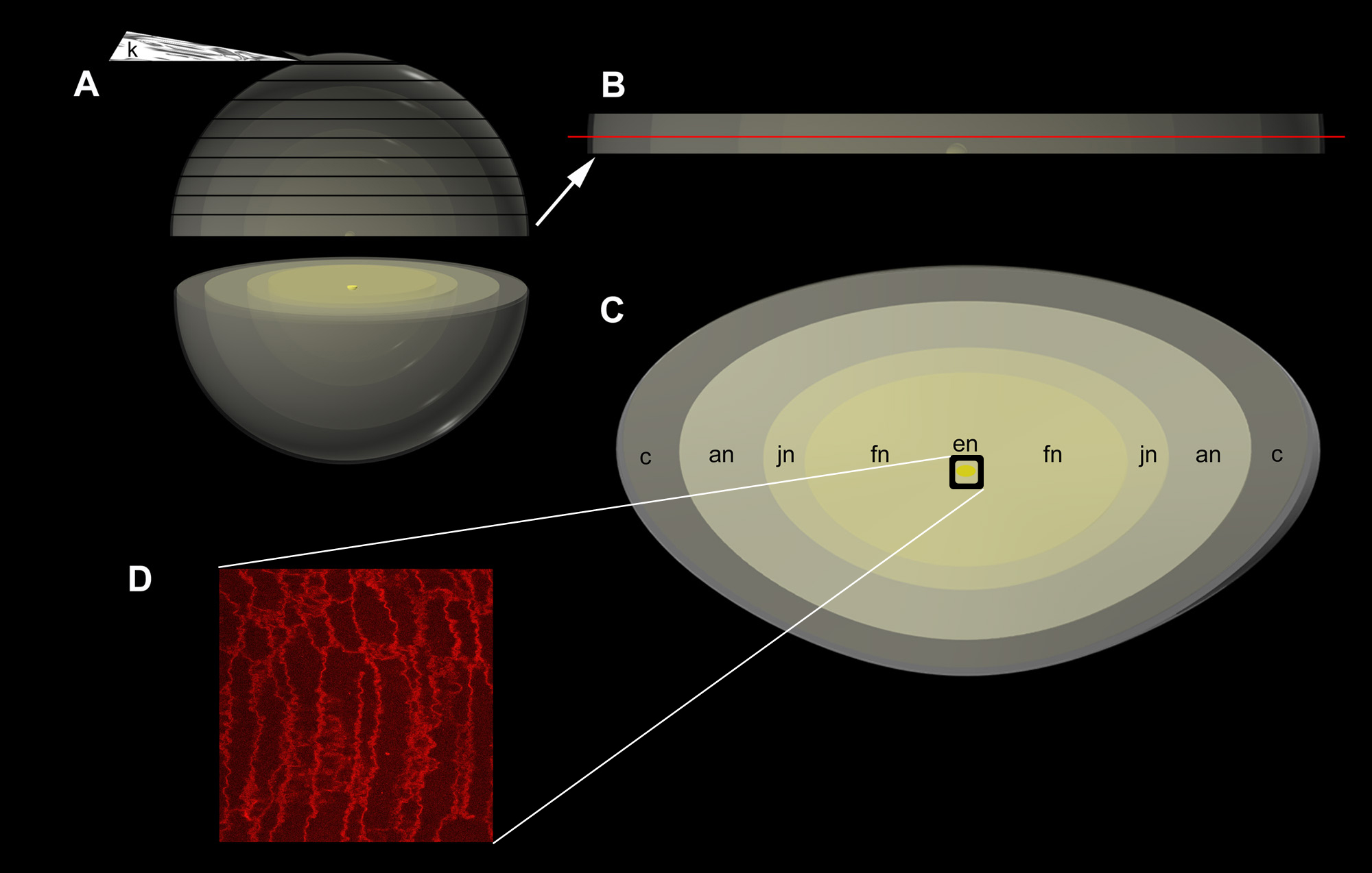
 Figure 1 of
Gilliland, Mol Vis 2008; 14:572-582.
Figure 1 of
Gilliland, Mol Vis 2008; 14:572-582.