Figure 3. Effect of homoisoflavanone on laser-photocoagulation-induced choroidal neovascularization. Choroidal neovascularization (CNV)
in control and homoisoflavanone-treated mice was evaluated by fluorescein angiography using 500,000 molecular weight fluorescein-conjugated
dextran. Wholemount preparation from control (A) and 1 µM homoisoflavanone-treated (B) mice subjected to laser-photocoagulation-induced CNV was performed after 1 h perfusion of fluorescein-conjugated dextran,
respectively. Circles indicate CNV in the laser-photocoagulation site. Hematoxylin-stained cross-sections were prepared from
control (C) and 1 μM homoisoflavanone-treated (D) mice subjected to laser, respectively. Arrows show the new vessels growing from choroidal vessels. E: To quantify CNV, we counted vessels from subretinal fibrovascular membrane. Data in each column are the mean ± standard
deviation values from 100 sites of 25 mice (*p<0.05). Scale bars in C and D equal 50 µm.
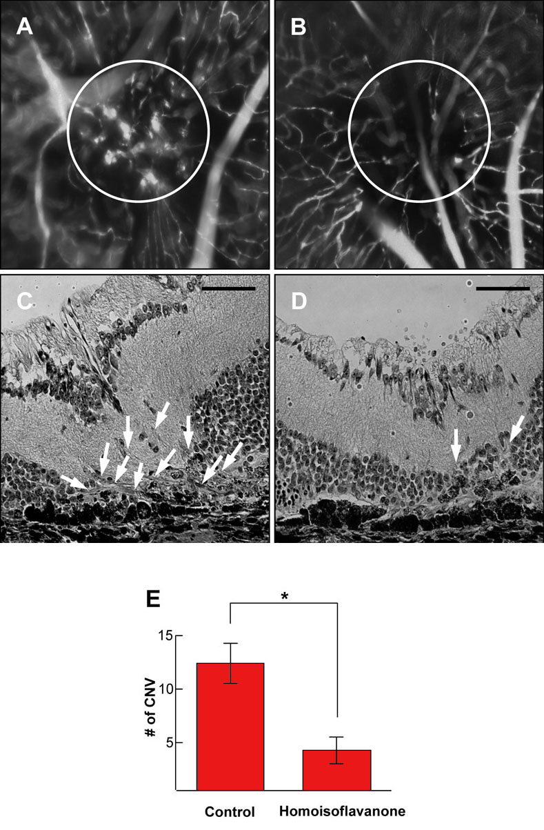
 Figure 3 of
Kim, Mol Vis 2008; 14:556-561.
Figure 3 of
Kim, Mol Vis 2008; 14:556-561.