Figure 6. Olomoucine treatment decreased
localization of MMP-2 protein at the wound edge. A: MMP-2
immunofluorescence is elevated at the wound edge in untreated control
corneas. B: The immunostaining of MMP-2 at the wound edge
(arrow) is reduced in olomoucine-treated corneas. C: Whole
mounted corneas without primary antibody showed no immunofluorescence. D:
Paraffin sections of wounded control corneas showed MMP-2
immunofluorescence in all layers of the cornea at wound edge (arrow)
and in basal epithelial cells distal to the wound (arrowhead). E:
The paraffin section of olomoucine-treated cornea showed reduced
immunofluorescence of MMP-2 at the wound edge (arrow). MMP-2
immunofluorescence persisted in the basal layers of epithelium
(arrowhead). F: No immunostaining was detected in the paraffin
section of the cornea when primary MMP-2 antibody was omitted. Scale
bar=100 μM.
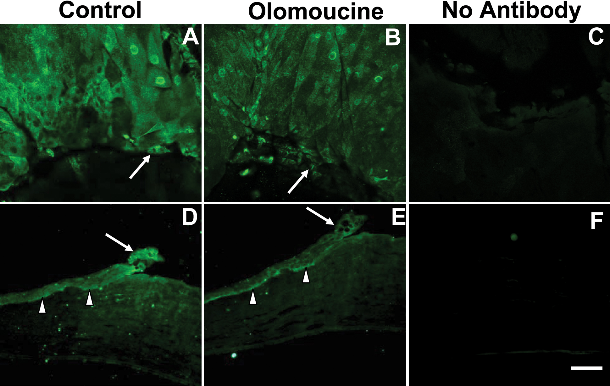
 Figure 6 of Tripathi, Mol Vis 2008; 14:542-549.