Figure 2. Distribution of ADORA1, ADORA2A, ADORA2B, and ADORA3 in HSF in vitro using indirect immunofluorescence. FITC-marked the secondary
antibody (green; 1) and PI dyed the nucleus (red; 2). (1) and (2) are combined into (3). ADORA1 is concentrated in the nucleus
of HSF (A1-A3). ADORA2A is mainly distributed on one side of the HSF cytoplasm (B1-B3). ADORA2B is distributed in the cytoplasm
and nucleus of HSF (C1-C3). ADORA3 is weakly expressed in the cytoplasm of HSF (D1-D3). Magnification: 400X.
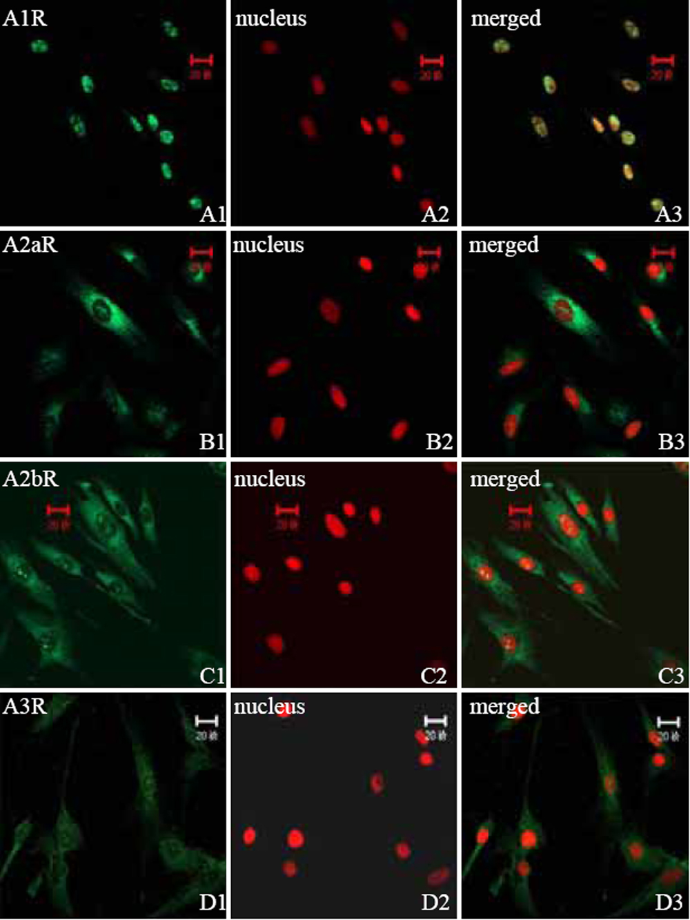
 Figure 2 of
Cui, Mol Vis 2008; 14:523-529.
Figure 2 of
Cui, Mol Vis 2008; 14:523-529.