Figure 1. Identification of HSF with vimentin, keratin, desmin, and S-100. PI dyed the nucleus (red) and the FITC-marked secondary antibody
(green). A: Vimentin antibody was added to primary cultured HSF migrating from a piece of sclera tissue. B: Negative controls used FITC-marked secondary antibodies of HSF. C: Vimentin is strongly expressed in the cytoplasm of HSF. D,E,F: Keratin, desmin, and S-100 protein are not expressed in the cytoplasm or nucleus of HSF. Magnification: 200X.
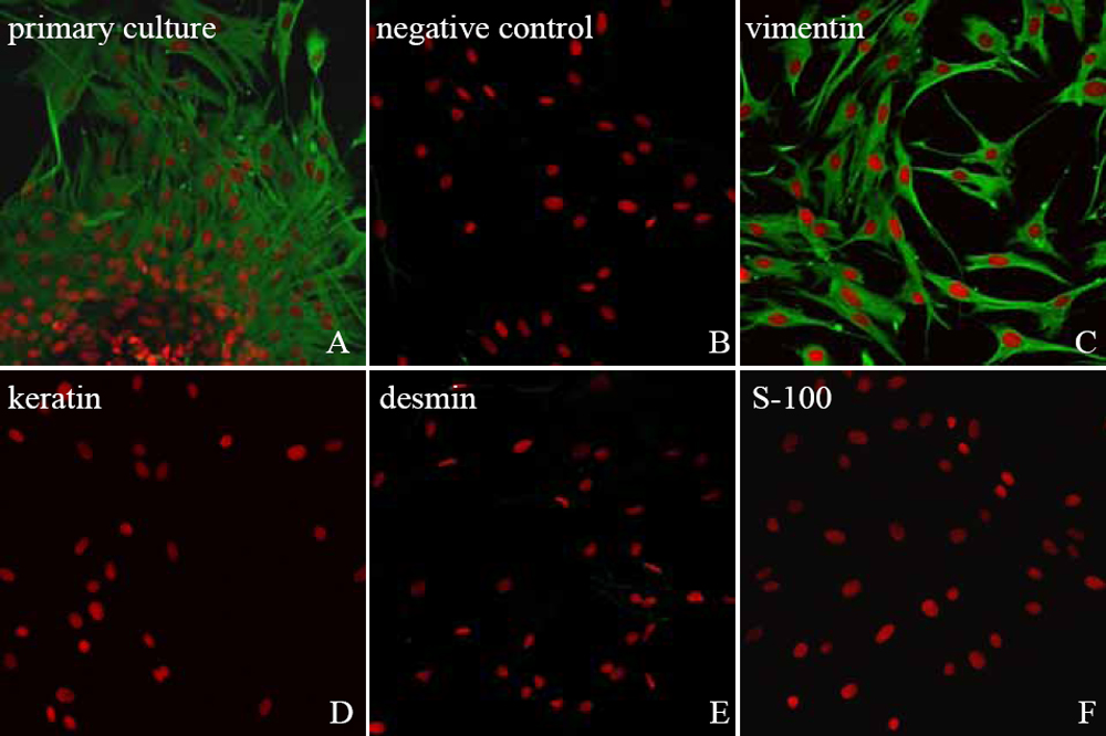
 Figure 1 of
Cui, Mol Vis 2008; 14:523-529.
Figure 1 of
Cui, Mol Vis 2008; 14:523-529.