Figure 1. Representative 2DE gel map of
tear proteins of normal subjects and fungal keratitis patients. Tear
proteins (270 μg) were separated using an 18 cm pH 3–10 NL IPG strip in
the first dimension and 12.5% SDS–PAGE followed by coomassie blue G-250
staining in the second dimension. The identified normal tear proteins,
cystatin S precursor (spot 1), cystatin SN precursor (spot 5), cystatin
(spot 29), human tear lipocalin (spot 49), prolactin inducible protein
(spot 34), serum albumin (spot 44), and fungal protein such as the
glutaredoxin-related protein (spot 54) are marked and compared between
control tear samples, Fusarium keratitis tear samples, and Aspergillus
keratitis tear samples.
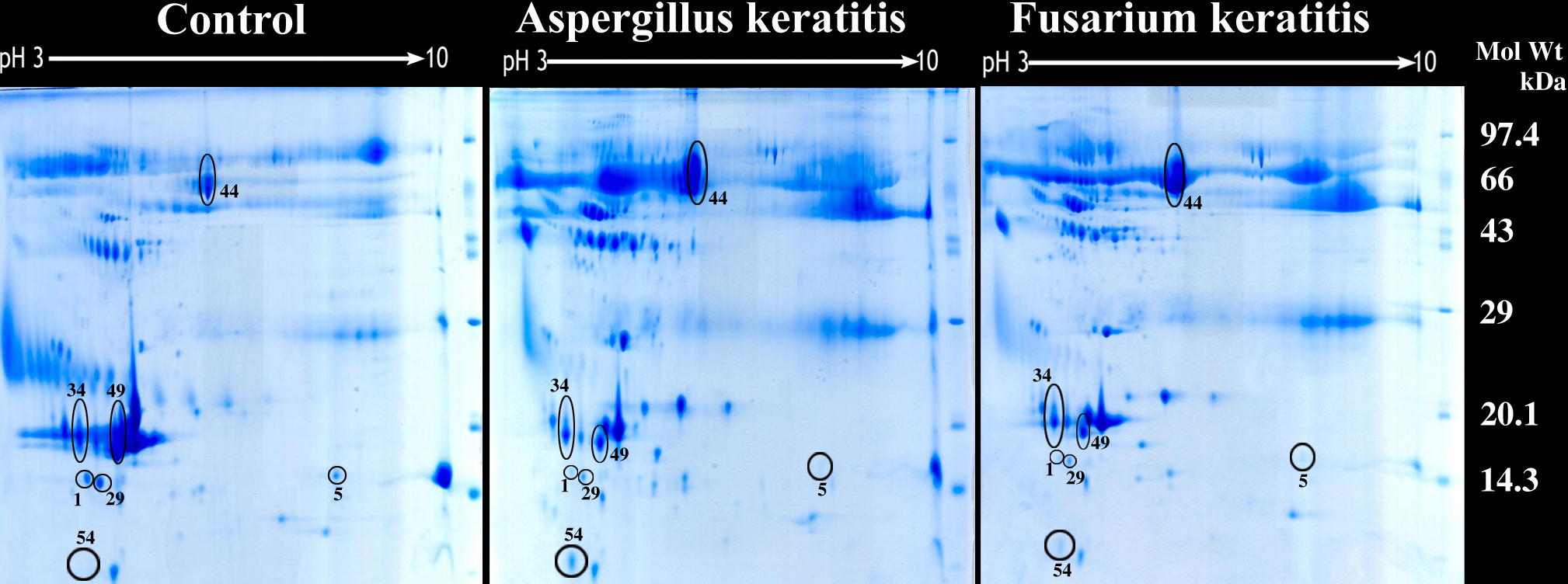
 Figure 1 of Ananthi, Mol Vis 2008; 14:500-507.