![]() Figure 2 of
Cools-Lartigue, Mol Vis 2008;
14:50-55.
Figure 2 of
Cools-Lartigue, Mol Vis 2008;
14:50-55.
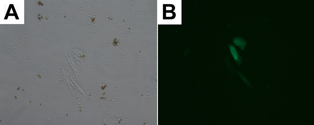
Figure 2. View of 92.1 cells isolated from spiked human blood
A: View at 10x magnification using conventional light microscopy of a colony of 92.1 transfected with GFP isolated from a sample of spiked human blood at a concentration of 10 cells/ml. B: View at 10x magnification using phase contrast microscopy of the colony of 92.1 transfected with GFP depicted in A isolated from a sample of spiked human blood at a concentration of 10 cells/ml.