![]() Figure 1 of
Cools-Lartigue, Mol Vis 2008;
14:50-55.
Figure 1 of
Cools-Lartigue, Mol Vis 2008;
14:50-55.
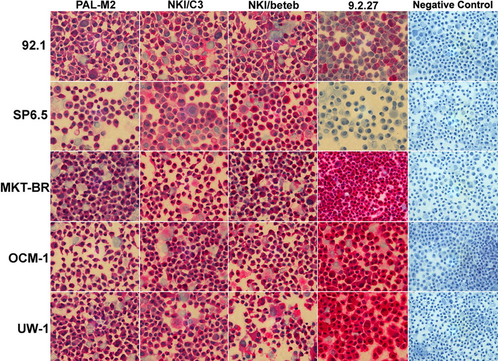
Figure 1. Immunohistochemical profile of 5 human uveal melanoma cell lines stained with monoclonal antibodies
Immunohistochemical profile of 5 human uveal melanoma cell lines (92.1, SP6.5, MKT-BR, OCM-1, and UW-1) stained with monoclonal antibodies PAL M2, NKI C3, NKI/Beteb, 9.2.27, and appropriate negative control. Each image is at 400x magnification.