Figure 1. Slit-lamp photographs of the
proband’s affected eye. A and B: Slit-lamp photographs
of the ropy lattice lines in the proband’s affected eye at two
different magnifications are shown. The proband was 47 years old. C
and D: Slit lamp photograph of the right and the left eye of
proband's brother showing multiple small geometric rod shaped lesions
in the posterior corneal stromal (the lesions are highlighted with
arrowheads).
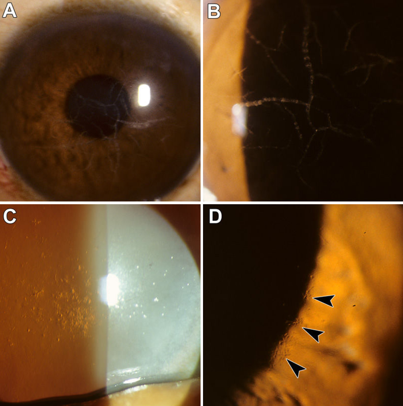
 Figure 1 of Afshari, Mol Vis 2008; 14:495-499.