Figure 1 of
Ramprasad, Mol Vis 2008; 14:481-486.
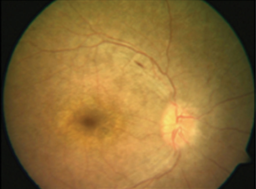
Figure 1.
Color fundus photograph of the right eye. Fundus photograph of the right eye of the proband showing midperipheral white dots at the level of the retinal pigment epithelium, arteriolar attenuation and an abnormal sheen in the macula.
 Figure 1 of
Ramprasad, Mol Vis 2008; 14:481-486.
Figure 1 of
Ramprasad, Mol Vis 2008; 14:481-486.