Figure 4. Demonstration of cell-specificity. A: Comparison of hypoxia induced dual luciferase activity of the HRSE-6xHRE-RPE65 promoter in retinal pigmented epithelium (ARPE-19)
cells with other cell types. The activation of the HRSE-6xHRE-Rpe65 promoter in ARPE-19 cells exposed to 1% oxygen for 40
hours was 51.3±10.8 (n=10, p<0.001) fold over aerobic activity. The non-epithelial cell lines displayed a relatively minimal
activation of the HRSE-6xHRE-Rpe65 promoter throughout the same hypoxic time course confirming a hypoxia-inducible and cell-specific
gene expression regulated by the HRSE-6xHRE-Rpe65 promoter construct. Hypoxia-mediated promoter activation for the tested
cell lines were: HT22 mouse hippocampal neuron (4.3±1.6, n=9, p<0.001), HEK-293 human embryonic kidney (3.3±2.5, n=6, p<0.001),
C6 glioma (2.8±0.3, n=5, p<0.001) and A-172 glioblastoma (1.2±1.4, n=3, p<0.001). B: The relative strength of the HRSE-6xHRE-Rpe65 promoter in hypoxia is comparable to cytomegalovirus (CMV). The histogram illustrates
that the HRSE-6xHRE-Rpe65 promoter activity was similar to the CMV promoter under hypoxic conditions. Under aerobic conditions
the CMV promoter activity remains strong while the HRSE-6xHRE-Rpe65 promoter is conditionally silenced. Asterisks (*) indicate
statistical significance.
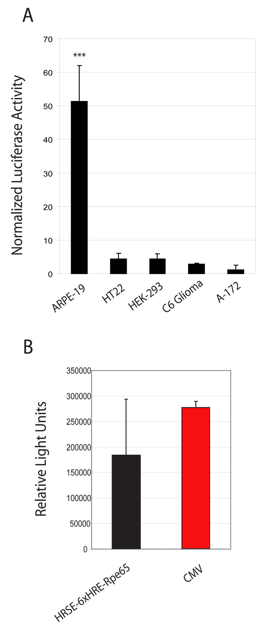
 Figure 4 of
Dougherty, Mol Vis 2008; 14:471-480.
Figure 4 of
Dougherty, Mol Vis 2008; 14:471-480.