Figure 6. 2D-SDS–PAGE. A seven centimeter
2D-SDS–PAGE of capillary collected and Schirmer extracted tear proteins
stained with SYPRO Ruby and overlaid to show the contrasting proteins
observed between the two collection methods. The red channel represents
the image for capillary collected tears and the blue channel represents
the image from the tear proteins extracted from a Schirmer strip.
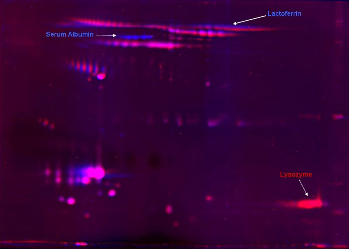
 Figure 6 of Green-Church, Mol Vis 2008; 14:456-470.