Figure 3. Comparison of collagen mRNA
levels in peripheral and central corneal tissues following
ovariohysterectomy. A, B, and C; Corneal
tissues were collected from a cohort of 54-week-old control and
ovariohysterectomized female rabbits. Corneal tissues were separated
into peripheral and central parts by using a 6 mm biopsy punch. mRNA
levels of collagen I, III, and V were determined by RT–PCR in these
tissues. Values are plotted as a percentage of Collagen I, Collagen
III, or Collagen V mRNA levels in normal control animals. Significant
changes compared with normal controls are indicated (p<0.05, p=not
significant [NS]).
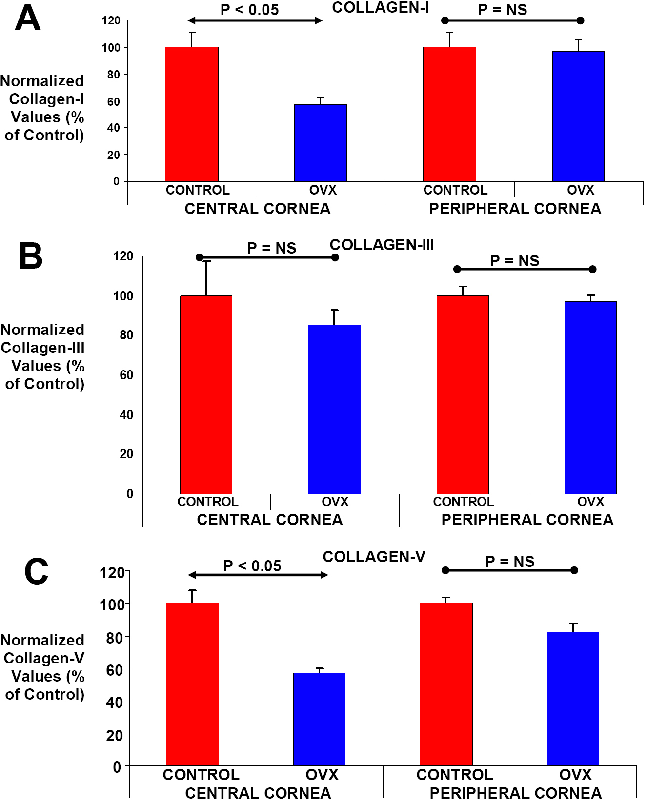
 Figure 3 of Achari, Mol Vis 2008; 14:443-455.