Figure 6. Epithelial phenotype of LEC and MC-L by Laser Scanning Confocal Microscopy and FACS analysis. LSCM pictures of limbal epithelial
cells (LECs) show positivity for K3 (20X) and K14 (20X; A,B), and LSCM pictures of MC-L show negativity for K3 (40X) and K14 (20X; C,D). The nuclei are counterstained with propidium iodide (blue, red). FACS histograms of MC-L confirm the absence of K3 (E) and K14 (F) expression. The blue line represents the test sample and the purple line represents the isotype control.
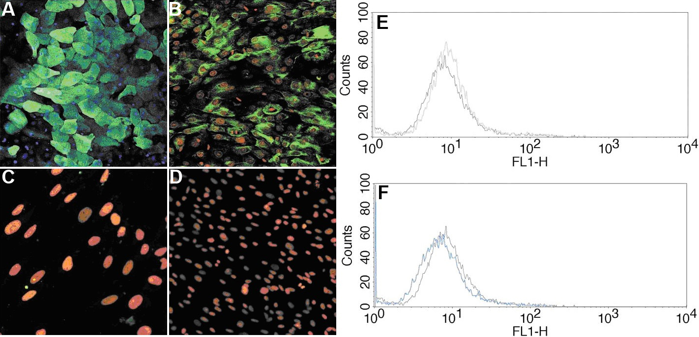
 Figure 6 of
Polisetty, Mol Vis 2008; 14:431-442.
Figure 6 of
Polisetty, Mol Vis 2008; 14:431-442.