Figure 4. Mesenchymal phenotype of MC-L by Laser Scanning Confocal Microscopy. LSCM pictures of MC-L show positivity (green fluorescence)
for vimentin (20X), CD90 (20X), and CD29 (20X) and negativity for CD11c (40X), CD11a (40X), CD45 (40X), CD34 (20X), and CD25
(20X). The nuclei are counterstained with propidium iodide (red).
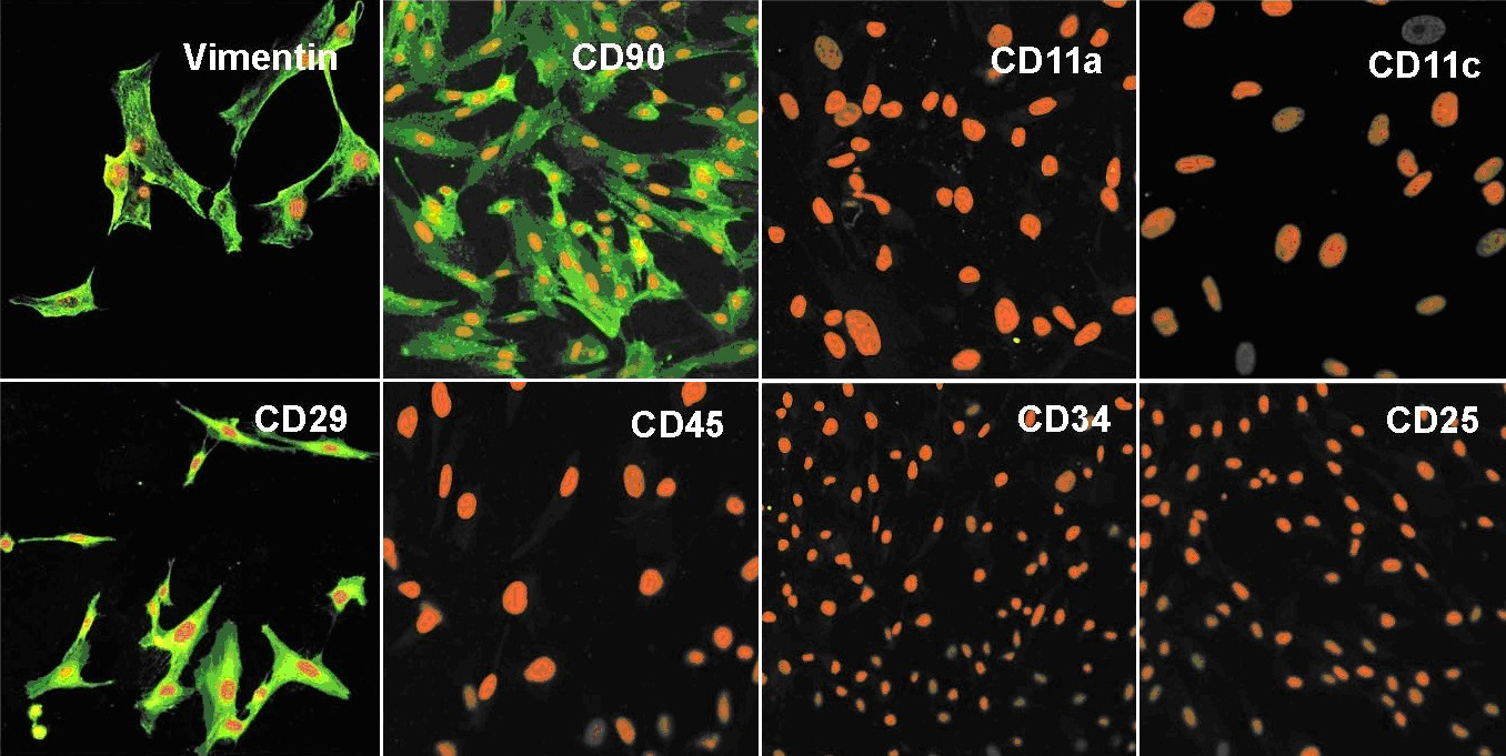
 Figure 4 of
Polisetty, Mol Vis 2008; 14:431-442.
Figure 4 of
Polisetty, Mol Vis 2008; 14:431-442.