Figure 2 of
Polisetty, Mol Vis 2008; 14:431-442.
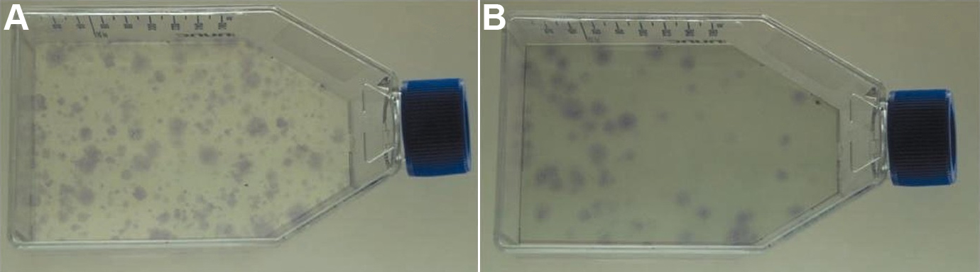
Figure 2.
Colony Formation Unit (CFU) assay of MC-L and MSC-BM. The figure shows the crystal violet stained colonies of stromal cells – MC-L (
A
) and MSC-BM (
B
) in T75 flasks.
 Figure 2 of
Polisetty, Mol Vis 2008; 14:431-442.
Figure 2 of
Polisetty, Mol Vis 2008; 14:431-442.