Figure 1. Morphology of cultured mesenchymal cells from limbus (MC-L) and mesenchymal stem or stromal cells from bone marrow (MSC-BM).
The phase contrast microscopic picture of MC-L shows the spindle morphology (magnification: 200X) (A); Cell sphere formation in the MC-L cultures gives the impression of embryoid body formation (magnification: 200X) (B); spindle shaped morphology of MC-L as confirmed by Giemsa stain (Light microscope, magnification: 200X) (C); Culture of MSC-BM (magnification: 200X) (D) showing spindle cell morphology similar to that of MC-L.
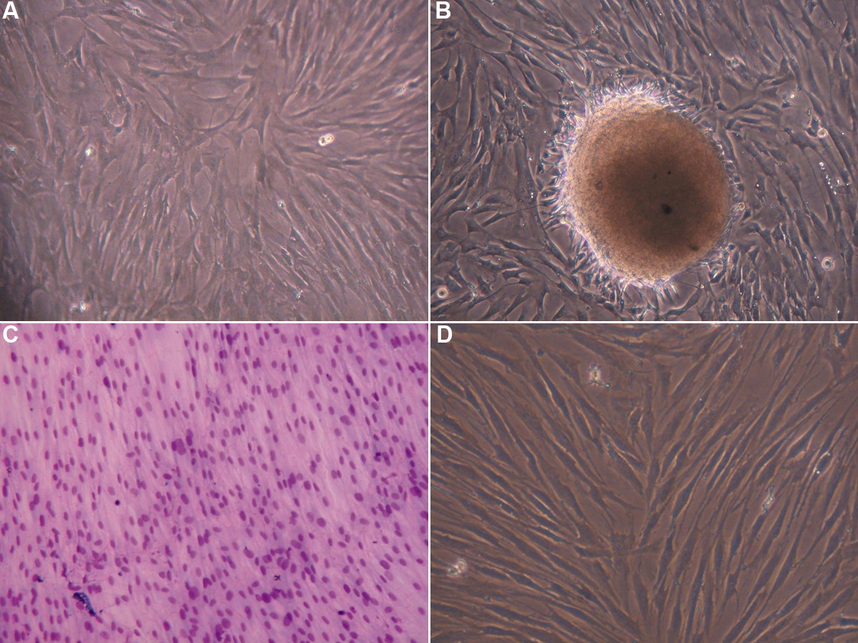
 Figure 1 of
Polisetty, Mol Vis 2008; 14:431-442.
Figure 1 of
Polisetty, Mol Vis 2008; 14:431-442.