Figure 3. Cross section of the lens of the proband’s mother (III: 2) under the scanning electron microscope. The normal lens tissue
was shown in a square while the opacity was shown in a circle (A). The parallel aligned strap fiber cells are represented in the longitudinal section (B). The structure of the opacities are puffy and irregularly aligned while floccules were observed and the fiber is tangled
(C,D).
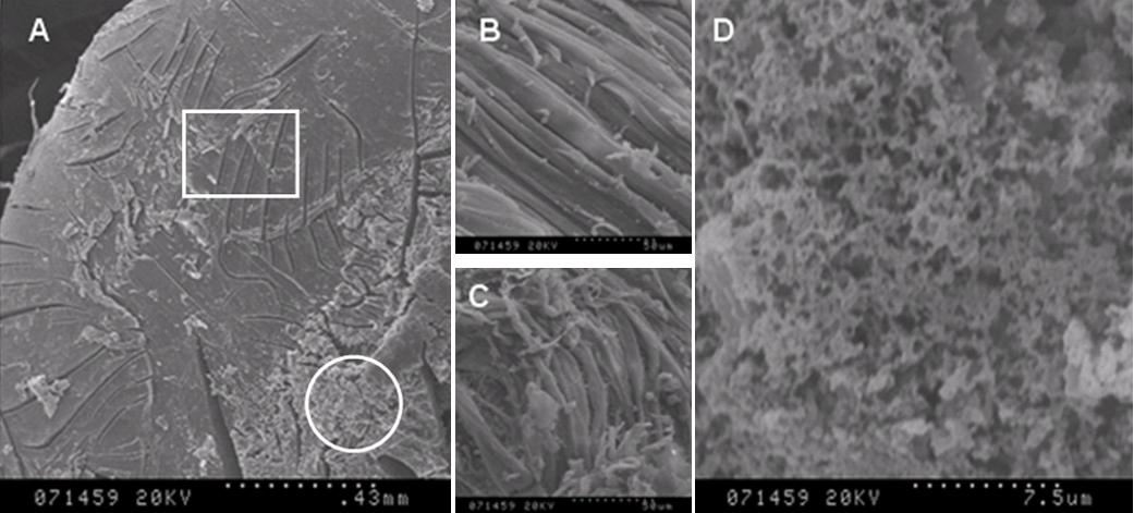
 Figure 3 of
Yan, Mol Vis 2008; 14:418-424.
Figure 3 of
Yan, Mol Vis 2008; 14:418-424.