![]() Figure 9 of
Zhang, Mol Vis 2008;
14:37-49.
Figure 9 of
Zhang, Mol Vis 2008;
14:37-49.
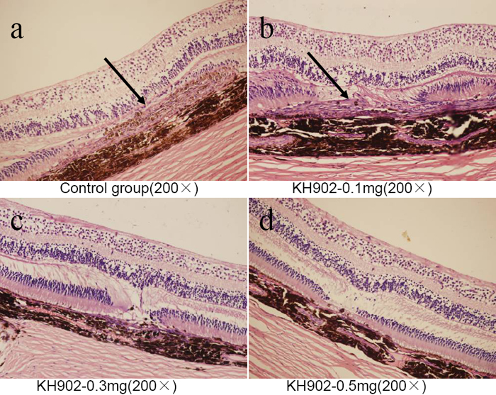
Figure 9. Ocular histology (hematoxylin-eosin stain) of all four groups at the endpoint of the experiment
The subretinal hyperblastosis (black arrowheads) in the control group and 100 μg KH902-treated group is shown. The outer nuclear layer was absent and retinal edema was obviously exhibited (A,B). In the 300 μg and 500 μg KH902-treated eyes, there were distinct retinal structures and no edema. The absent outer nuclear layer and a few fibrocytes were observed (C,D).