![]() Figure 4 of
Zhang, Mol Vis 2008;
14:37-49.
Figure 4 of
Zhang, Mol Vis 2008;
14:37-49.
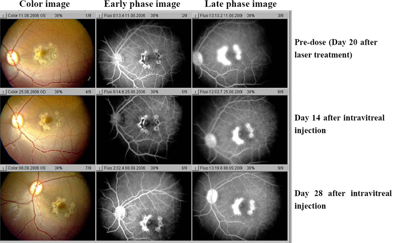
Figure 4. Color photography and angiography of an eye that received injection of vehicle taken on day 20 after laser and on days 14 and 28 after intravitreal injection
Color photo and fluorescein angiography of experimental model of choroidal neovascularization treated with injection of vehicle is shown. Note the gray-white change of lesions, local retina edema on color image and the large confluent bridging lesions and extensive late leakage on angiography image in left eye. There were no changes among three time points: day 20 after laser, day 14 and 28 after intravitreal injection.