![]() Figure 10 of
Zhang, Mol Vis 2008;
14:37-49.
Figure 10 of
Zhang, Mol Vis 2008;
14:37-49.
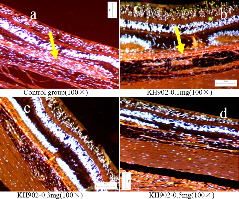
Figure 10. Merged images of immunohistochemistry
Immunohistochemistry analysis of proliferation-associated endothelial cells (stained by CD31/CD105 and DAPI) in all four groups at the endpoint of the experiment is shown. The proliferation-associated endothelial cells of choroidal neovascularization were stained with CD31 and CD105 (yellow arrowheads) like choroidal vessel endothelial cells in the control eye and 100 μg KH902-treated eye (A,B) whereas no positive stained proliferation-associated endothelial cells were found in 300 μg and 500 μg KH902-treated eye (C,D).