Figure 8. Standard immunofluorescence staining on Chang (WKD) and IOBA-NHC cells. Phalloidin and propidium iodide stainings show the
morphology of Chang cells (A,B) and IOBA-NHC cells (C,D) after 15 min of treatment with solutions of PBS (A,C) and BAC 10−2% (B,D). Nuclei were stained in red by propidium iodide and cytoskeleton (F-actin) in green by phalloidin. Note the BAC-dependent
increase of cell shrinkage on both cell lines. BAC, benzalkonium chloride; PBS, phosphate buffered saline.
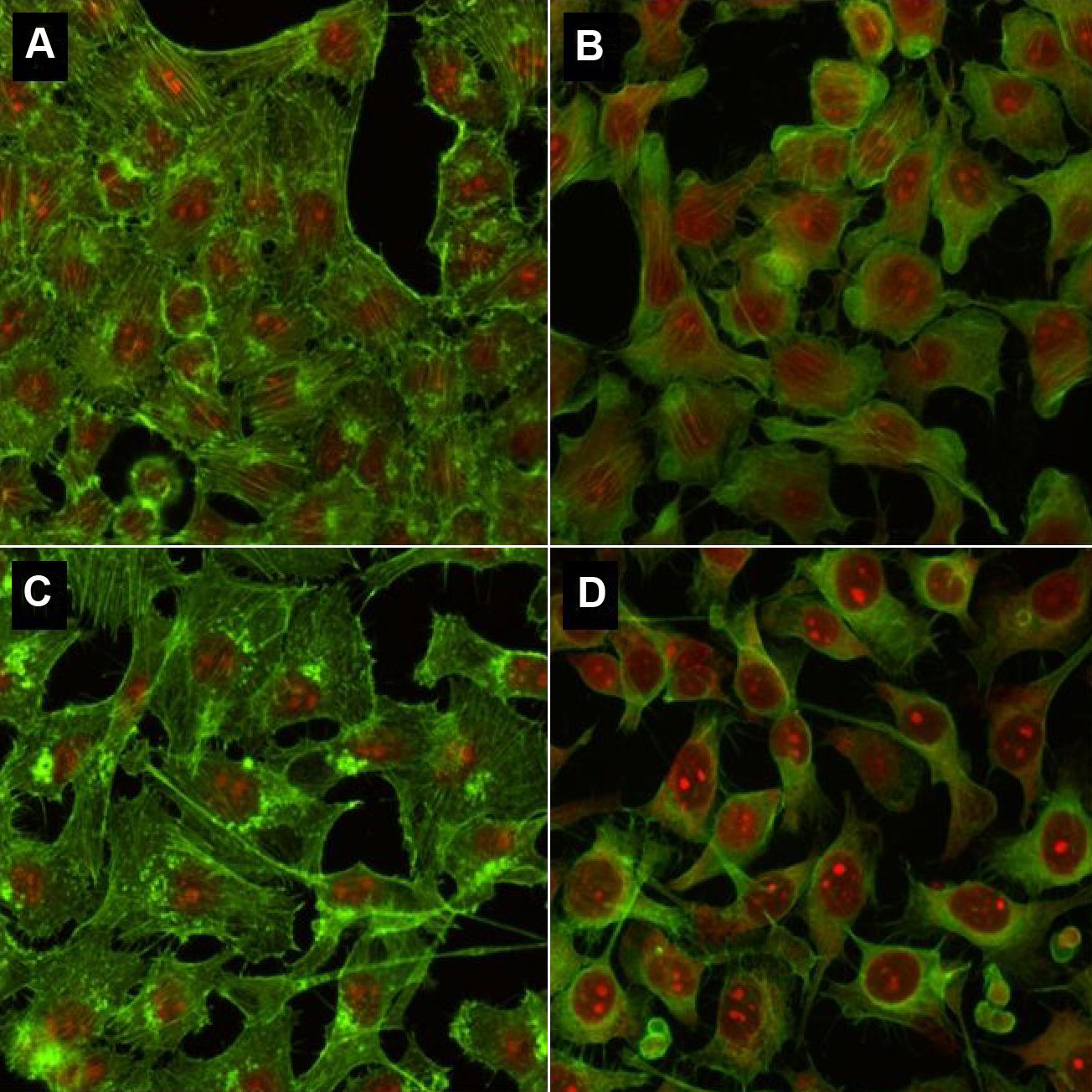
 Figure 8 of
Brasnu, Mol Vis 2008; 14:394-402.
Figure 8 of
Brasnu, Mol Vis 2008; 14:394-402.