Figure 4. Cellular localization of mRNA of the candidate genes in light-adapted carp retina. Cellular localization of some of the candidate
genes was determined with the in situ hybridization (ISH) method. A and B: ISH signals of rod candidate genes. A: Cyclic nucleotide gated channel (r1). B: O-GlcNAc transferase (r4). C-F: ISH signals of cone candidate genes. C: N-myc downstream regulated gene 1-like (c1). D: Aryl hydrocarbon receptor 2 (c2). E: Acyl CoA binding domain containing (c3). F: 14–3–3 Theta polypeptide (c4). G-L: Negative controls of A-F with use of sense probes. The following abbreviations were used: outer segment layer (OS), outer
nuclear layer (ONL), outer plexiform layer (OPL), inner nuclear layer (INL), inner plexiform layer (IPL), and ganglion cell
layer (GCL).
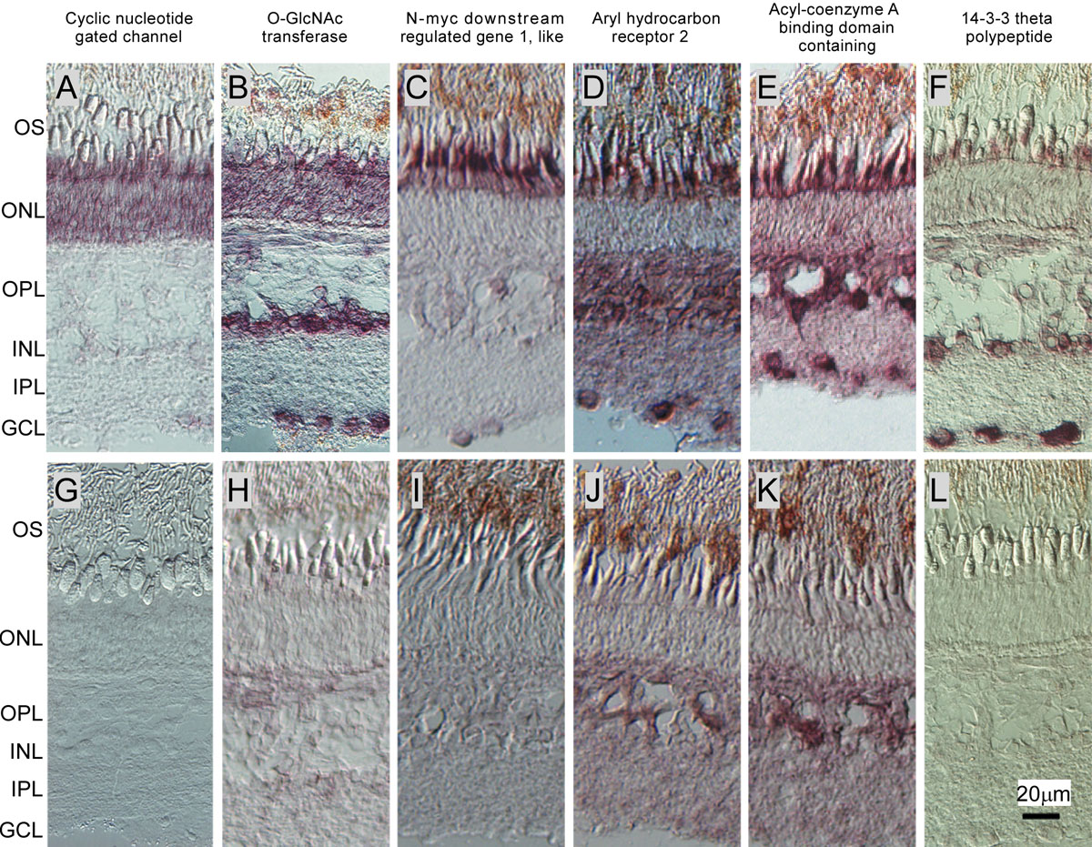
 Figure 4 of
Shimauchi-Matsukawa, Mol Vis 2008; 14:358-369.
Figure 4 of
Shimauchi-Matsukawa, Mol Vis 2008; 14:358-369.